Automatic eye fundus image vessel detecting method based on PCNN (pulse coupled neural network)

A fundus image, automatic detection technology, applied in the field of medical diagnosis, can solve problems such as low contrast between blood vessels and background

Inactive Publication Date: 2013-03-27
TIANJIN POLYTECHNIC UNIV
View PDF0 Cites 34 Cited by
  • Summary
  • Abstract
  • Description
  • Claims
  • Application Information

AI Technical Summary

Problems solved by technology

[0006] The present invention adopts the method based on self-adaptive PCNN, considers the problem that the contrast ratio of blood vessel and background in the actual fundus image is low, first uses the adaptive histogram equalization (Contrast Limited Adaptive HistogramEqualization, CLAHE) of contrast limitation and two-dimensional Gaussian matched filter Process the fundus image to enhance the contrast of blood vessels and background; then based on the simplified PCNN model, use the Energy of Laplace (EOL) of the pixel as the link strength value of the corresponding PCNN neuron, combined with the maximum inter-class variance The fundus image is segmented according to the criteria; finally, the final blood vessel detection result is obtained by area filtering

Method used


Image

  • Automatic eye fundus image vessel detecting method based on PCNN (pulse coupled neural network)
  • Automatic eye fundus image vessel detecting method based on PCNN (pulse coupled neural network)
  • Automatic eye fundus image vessel detecting method based on PCNN (pulse coupled neural network)

Examples


Embodiment Construction

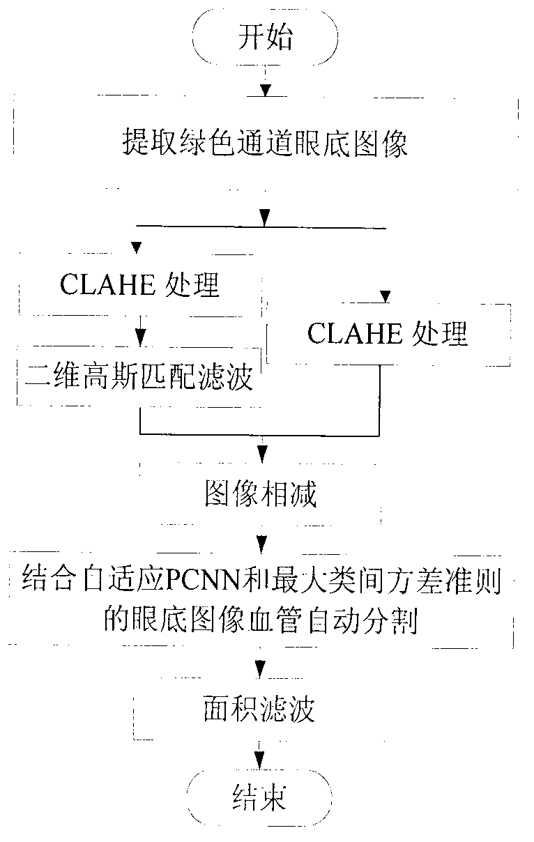
[0065] The flow chart of the present invention is as figure 1 As shown, the fundus image of the green channel with high contrast between blood vessels and the background is first extracted, and the fundus image is processed by combining CLAHE and two-dimensional Gaussian matching filter, and then based on the simplified PCNN model, the EOL of the pixel is used as the corresponding PCNN neuron The link strength value of the method is combined with the maximum between-class variance criterion to segment the fundus image, and finally the final blood vessel detection result is obtained by area filtering. The specific implementation process of the technical solution of the present invention will be described below in conjunction with the accompanying drawings.

[0066] 1. Fundus image preprocessing:

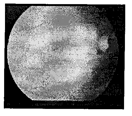
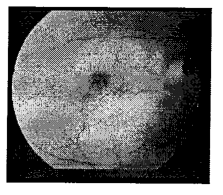
[0067] 1.1 First use CLAHE to process the fundus image of the green channel, Figure 4 for right image 3 Fundus image after performing CLAHE.

[0068] 1.2 For the CLAHE processin...


PUM


Abstract

The invention relates to an automatic eye fundus image vessel detecting method based on a PCNN (pulse coupled neural network). CLAHE (contrast limited adaptive histogram equalization) with limited contrast ratio and two-dimensional Gaussian matched filtering are used for processing an eye fundus image, pixel EOL (energy of Laplace) is used, on the basis of simplified PCNN models, as link intensity value of corresponding PCNN neurons, the eye fundus image is then cut according to the maximum interclass variance standard, and final vessel detecting results are obtained through area filtering. By fully utilizing advantages of combination of the PCNN and the method, the vessel network of the eye fundus image can be fully and accurately detected.

Description

technical field [0001] The invention relates to an automatic detection method of blood vessels in fundus images based on adaptive PCNN. The method can extract complete main blood vessels and tiny blood vessels from normal and lesioned fundus images, belongs to the technical field of image processing, and can be applied to medicine diagnosis. Background technique [0002] The eye is the most important visual organ of the human body, and the fundus blood vessels are the only deep microvessels in the human body that can be directly observed in a non-invasive way. Compared with other tissues, fundus blood vessels are the most stable and obvious in fundus images. Many systemic diseases, such as diabetes, hypertension, arteriosclerosis and other cardiovascular and cerebrovascular diseases, will affect the brightness, position and shape of fundus blood vessels. Therefore, the detection of blood vessels in fundus images is an important research topic. Diagnosis has important guidi...

Claims


Application Information

Patent Timeline
Patent Type & Authority Applications(China)
IPC IPC(8): G06T7/00
Inventor 吴骏肖志涛耿磊张芳王淑芹
Owner TIANJIN POLYTECHNIC UNIV
Why Eureka
Try Eureka