Perfusion fixation device and method for small animal experiment

A technology for fixing devices and small animals, used in medical science, veterinary instruments, etc., can solve problems such as troublesome operation, low pressure, tissue damage, etc., and achieve the effect of good effect, time-saving and labor-saving work efficiency.

Inactive Publication Date: 2013-05-01
ARMY MEDICAL UNIV
View PDF9 Cites 11 Cited by
  • Summary
  • Abstract
  • Description
  • Claims
  • Application Information

AI Technical Summary

Problems solved by technology

[0006] (1) Manual control of the syringe is not easy to control the pressure: when the perfusion fixation method uses a simple device combined with a syringe and an injection needle, the pressure is manually controlled by the operator. If the pressure is too small, the perfusion effect will not be good; Damage (eg, burst blood vessel, burst heart)
When using a simple device combined with an infusion bottle and an injection needle, it is suitable for rats and rabbits, but for small animals, the pressure of the device is obviously too small due to the small blood vessels, and the perfusion effect cannot be ensured.
[0007] (2) At least two people are required to operate the simple device at the same time: when using a simple device combined with a syringe and an injection needle, two or more people are usually required to cooperate at the same time, and one person is required to complete the anatomical perfusion and control the pressure of the syringe, and the operation is more difficult. Trouble, may lead to incomplete brain tissue structure of small animals, affecting the authenticity, reliability and repeatability of experimental results

Method used


Image

  • Perfusion fixation device and method for small animal experiment
  • Perfusion fixation device and method for small animal experiment
  • Perfusion fixation device and method for small animal experiment

Examples


Embodiment Construction

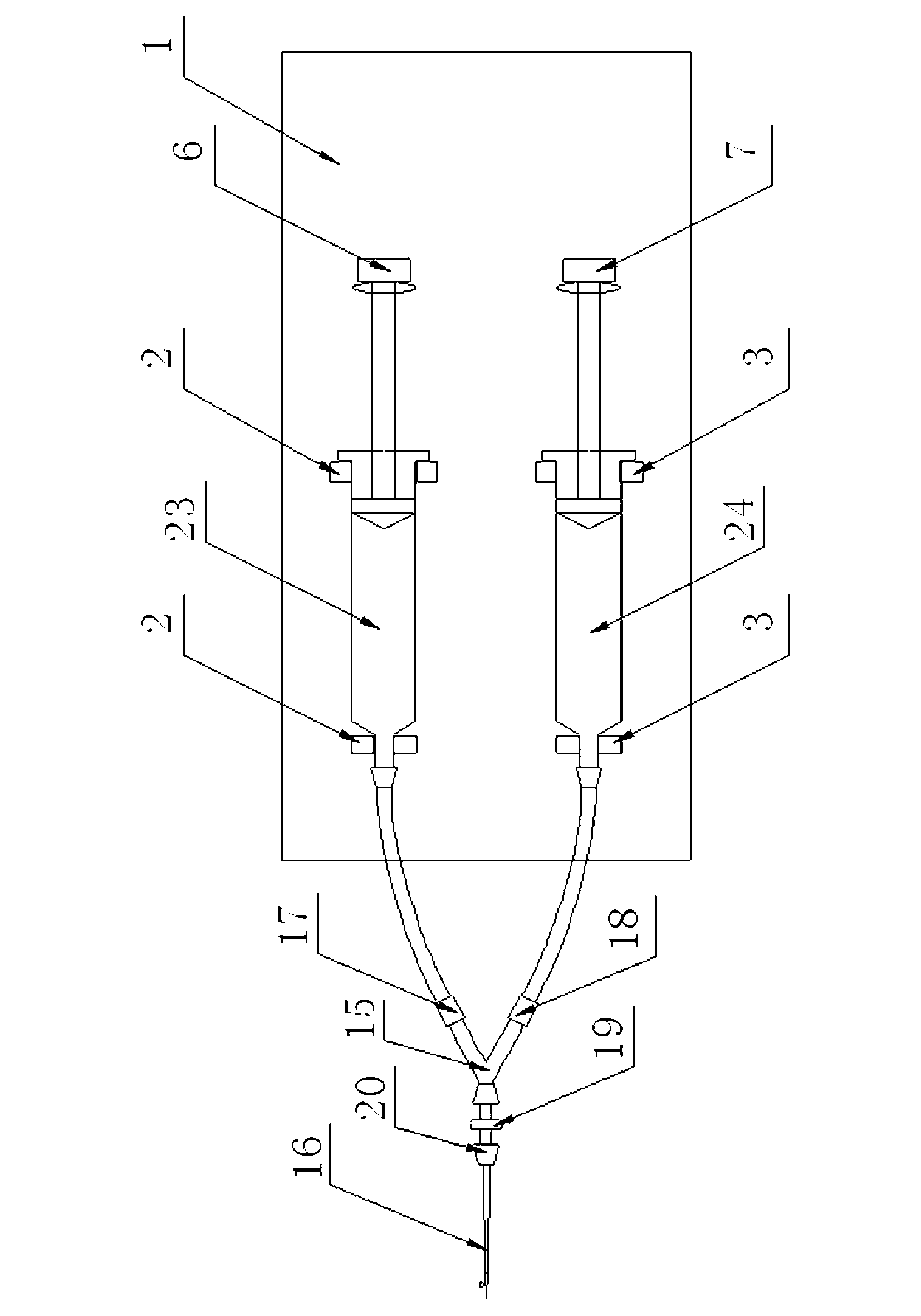
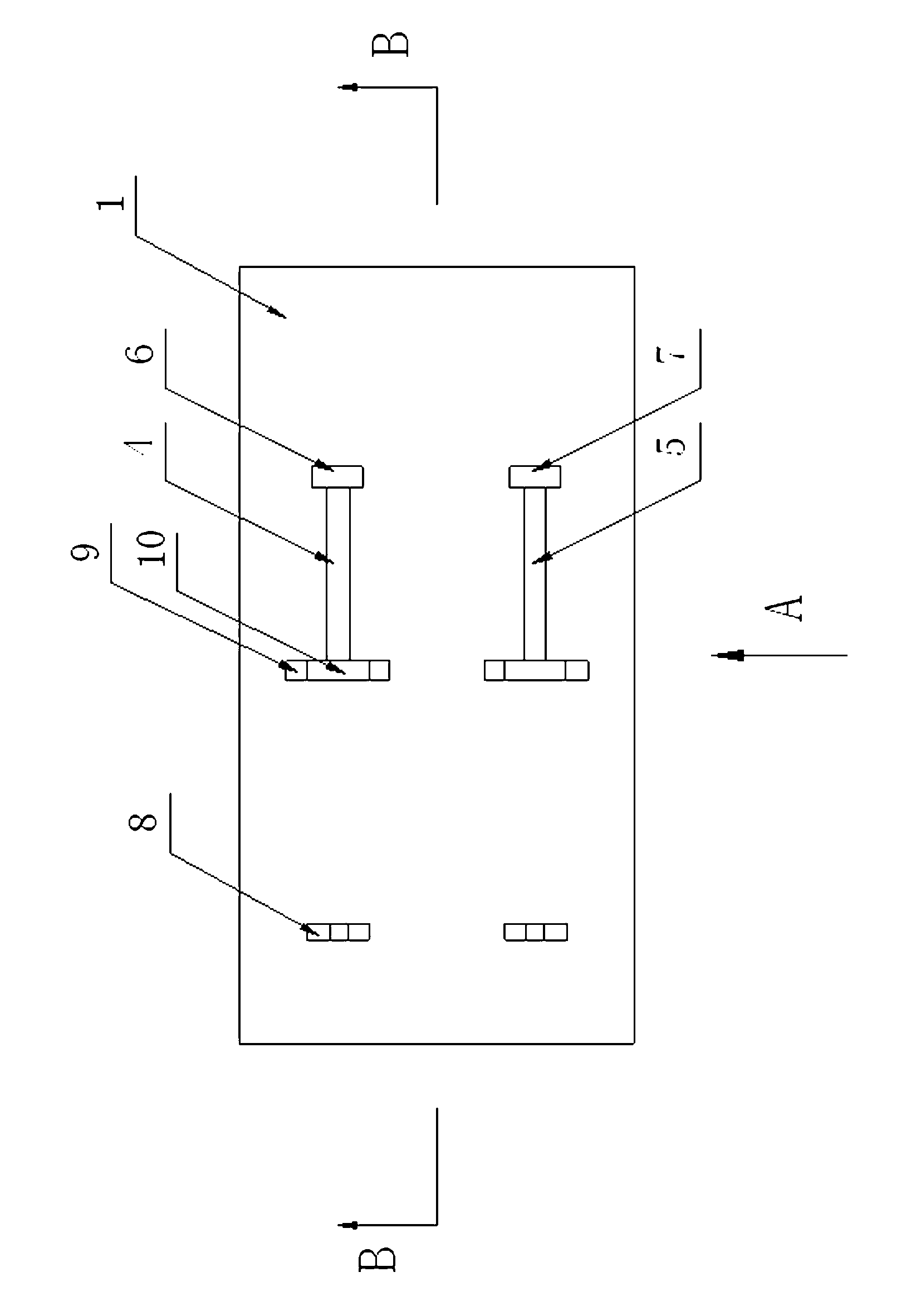
[0037] See Figure 1 to Figure 5 , Is an embodiment of the perfusion fixation device for the small animal experiment of the present invention. A perfusion fixing device for a small animal experiment, comprising a base 1. The upper end of the base 1 is provided with a first syringe fixing seat 2 and a second syringe fixing seat 3, and a first guide chute 4 is fixed to the first syringe The seat 2 corresponds to a second guide chute 5 corresponding to the second syringe holder 3. The first guide chute 4 is slidably fitted with a first slide 6 and the second guide chute 5 is slidably fitted with a second slide Block 7. The first syringe holder 2 and the second syringe holder 3 both include a front holder 8 and a rear holder 9. The upper ends of the front holder 8 and the rear holder 9 of the first syringe holder 2 are both provided with positioning The upper ends of the groove 10, the front fixing seat 8 and the rear fixing seat 9 of the second syringe fixing seat 3 are all prov...


PUM


Abstract

The invention relates to a perfusion fixation device and a perfusion fixation method for a small animal experiment. The device comprises a pedestal, wherein the upper end face of the pedestal is provided with a first syringe fixing seat, a second syringe fixing seat, a first guide sliding chute and a second guide sliding chute; the first guide sliding chute corresponds to the first syringe fixing seat; the second guide sliding chute corresponds to the second syringe fixing seat; a first slide block is arranged in the first guide sliding chute in a sliding fit manner; a second slide block is arranged in the second guide sliding chute in a sliding fit manner; a first step motor and a second step motor are arranged in the pedestal respectively; the first step motor is connected with the first slide block through a first lead screw nut mechanism; the second step motor is connected with the second slide block through a second lead screw nut mechanism; both the first step motor and the second step motor are electrically connected with a motor control circuit; the pedestal is provided with a perfusion flow speed adjusting key and a motor control switch; and the perfusion flow speed adjusting key and the motor control switch are both electrically connected with the motor control circuit. The device and the method have the advantages of good perfusion effect, convenience in operation and simple structure.

Description

Technical field [0001] The invention relates to the technical field of perfusion fixation of experimental animals, in particular to a perfusion fixation device and method for small animal experiments. Background technique [0002] In experimental animal histopathology research, especially in situ hybridization and immunohistochemistry research, in order to avoid or reduce the changes in tissue morphology and structure after death, perfusion fixation is often used before the animal is sacrificed. Perfusion fixation is to introduce the fixative into the tissues and organs that need to be fixed through the vascular route, and quickly fix the living cells in situ in time, and then take the sample. The tissue fixed by perfusion has a certain degree of hardness, and all its tissue structures are maintained in the pre-mortem state. Therefore, it is of great significance to choose the tissues fixed by perfusion to maintain the integrity of the morphology and tissue structure. [0003] The...

Claims


Application Information

Patent Timeline
Patent Type & Authority Applications(China)
IPC IPC(8): A61D7/00
Inventor 杨萍邓玲张明伟温惠中左峰秦余
Owner ARMY MEDICAL UNIV
Why Eureka
Try Eureka