
Method for rapidly concentrating and extracting nucleic acid target cells from sample

A technology of cell nucleic acid and target cells, applied in the field of nucleic acid extraction, to achieve the effect of increasing the positive rate, increasing the concentration of nucleic acid, and good social effects

Inactive Publication Date: 2013-07-31
南京紫霄科技有限公司 +2
View PDF0 Cites 7 Cited by
  • Summary
  • Abstract
  • Description
  • Claims
  • Application Information

AI Technical Summary

Problems solved by technology

[0004] At present, there are a few technologies for extracting nucleic acid from paraffin tissue in China. Basically, no nucleic acid is extracted after cutting white slices and then enriching and protecting the dewaxed slices by HE staining. Instead, the target cell area of ​​the specimen is observed without HE staining, and the target cell area is mechanically broken. Or the method of directly dewaxing in the centrifuge tube after boiling the paraffin tissue or rolling film

Method used


Image

  • Method for rapidly concentrating and extracting nucleic acid target cells from sample
  • Method for rapidly concentrating and extracting nucleic acid target cells from sample
  • Method for rapidly concentrating and extracting nucleic acid target cells from sample

Examples


Embodiment 1

[0033] The dewaxing of embodiment 1 white sheet

[0034] Select several white sheets (2-15 sheets) depending on the size of the specimen and soak them in xylene for 8-12 hours to fully dissolve the paraffin; replace with new xylene to dissolve the paraffin and soak for 15-20 minutes; replace xylene again and soak for 15 minutes; Immediately soak the slices in absolute ethanol for more than 10 minutes to fully dissolve xylene; replace with new absolute ethanol, soak for 10 minutes, and then soak in 95% ethanol for 5 minutes; 80% ethanol for 5 minutes; 70% ethanol for 2 minutes; tap water for a while 2min, washed with distilled water for 2min, blotted dry, and then added dropwise 50uL of nucleic acid protection solution. Under the microscope, select the target cells on the HE stained sheet with a marker pen, and under the guidance of the HE sheet, also select the target cells with a marker pen at the same position on the white sheet, scrape off the target cells, enrich them, and...

Embodiment 2

[0036] Example 2 Selection of target cells under the guidance of HE-stained slices of colorectal cancer (pathological number 082379)

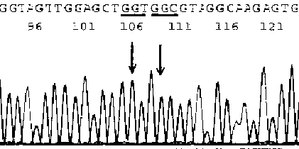
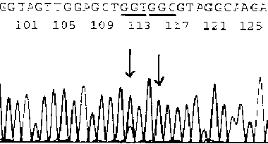
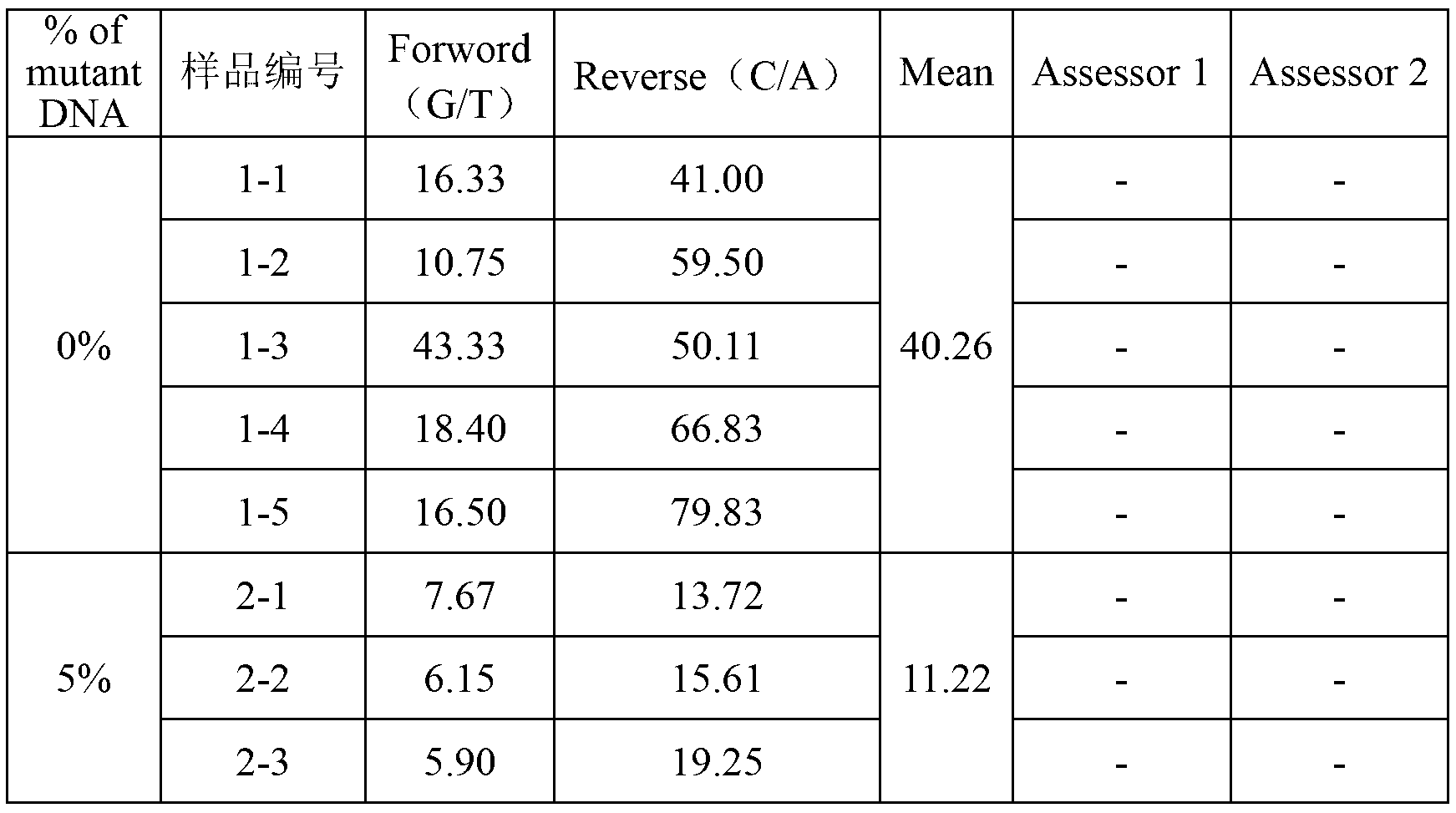
[0037]HE-stained sections must be re-examined and diagnosed by pathologists or cytologists before nucleic acid extraction to confirm that they contain target cell components, and mark the location of target cells on the slide. Using the method of Example 1, extract the DNA template, and use the primers of the K-ras-2 gene (K-ras-2-U: -aggcctgctgaatttgactg-, K-ras-2-L: -catgattttggtcagagaaacc-;) for PCR amplification (QIAGEN HotStar DNA polymerase Kit10×amplification buffer 2uL, dNTP2U, upstream and downstream primers 10pmol each, template DNA 0.1~2ug Taq DNA polymerase 2.5u, Mg 2+ 1.5mmol / L add double distilled water to 20ul. PCR thermal cycle conditions: 95°C for 10min, 95°C for 40s, 60°C for 1min, 72°C for 45s, 30 cycles, 72°C for 45min, 4°C for incubation), the product was purified and sequenced (conventional method), the results are as f...


PUM

PropertyMeasurementUnit
melting pointaaaaaaaaaa
thicknessaaaaaaaaaa

Abstract

The present invention discloses a method for rapidly concentrating and extracting nucleic acid target cells from a sample, wherein the method comprises the steps of fixing surgical or biopsy specimens with neutral formalin, dehydrating to be embedded in paraffin, cutting multiple white slices and selecting one for HE staining, observing tumor tissue cell distribution of HE stained slices under an optical microscope, and marking the distribution area of target cells; baking the remained white slices for xylene dewaxing, scraping the tissue rich in target cells on the white slices according to the marked HE stained slices for target cells concentrating, and extracting nucleic acid from the concentrated target cells. The method can improve concentration of nucleic acid in the target cells to up to more than 40% to 60% so as to raise positive rate of molecular pathology detection and reduce false negative rate without any expensive equipment or specialized complex technology with almost no obvious time consumption, therefore the method is suitable for being applied in all inspection department of pathology and biological laboratories and is used to provide more accurate molecular diagnosis for clinical diagnosis and treatment.

Description

technical field [0001] The invention relates to a method for extracting nucleic acid, in particular to a method for rapidly enriching and extracting target cell nucleic acid from a sample. Background technique [0002] The existing method for extracting nucleic acid from paraffin tissue sections first uses a paraffin microtome to dewax the thin tissue cells contained in paraffin tissue block sections (also known as cut white slices or sliced ​​slices), dissolves alcohol into water, and then performs protease digestion , after refrigerated centrifugation and purification, use the phenol chloride method or kit to extract nucleic acid. Since the tissue cells with different benign and malignant properties co-grow in the sliced ​​tissue exist in different proportions and are different, the total nucleic acid composition of the extracted cells is a mixture of the above-mentioned tissue cells with different properties, which affects the authenticity of the target data. In order to...

Claims


Application Information

Patent Timeline
Patent Type & Authority Applications(China)
IPC IPC(8): C12N15/10
Inventor 赖仁胜吴晓斌谢玲陈劼余波赵明孙军华张树鹏
Owner 南京紫霄科技有限公司
Learn More