
Nonparametric automatic detection method of focal niduses

An automatic detection, non-parametric technology, used in instruments, character and pattern recognition, computer parts and other directions, can solve the problems of reducing the identification ability of local image descriptors, overfitting of parameter models, poor data classification performance, etc. Convenience, stable class estimation, and the effect of accurate detection

Active Publication Date: 2013-10-23
SOUTHERN MEDICAL UNIVERSITY
View PDF7 Cites 8 Cited by
  • Summary
  • Abstract
  • Description
  • Claims
  • Application Information

AI Technical Summary

Problems solved by technology

This method of lesion detection and segmentation based on a parametric model has several shortcomings: first, for different training samples (such as images of different lesion types), this method needs to retrain the parameters in the model, which is not easy; second, Since the training samples are generally relatively small, the parameter model will produce an overfitting phenomenon, that is, the classification judgment can be performed well for the training samples, but the classification performance for the data other than the sample set is poor.
The bag of words model needs to quantify the image local descriptors, and quantization will reduce the discriminative ability of the image local descriptors

Method used


Image

  • Nonparametric automatic detection method of focal niduses
  • Nonparametric automatic detection method of focal niduses

Examples


Embodiment 1

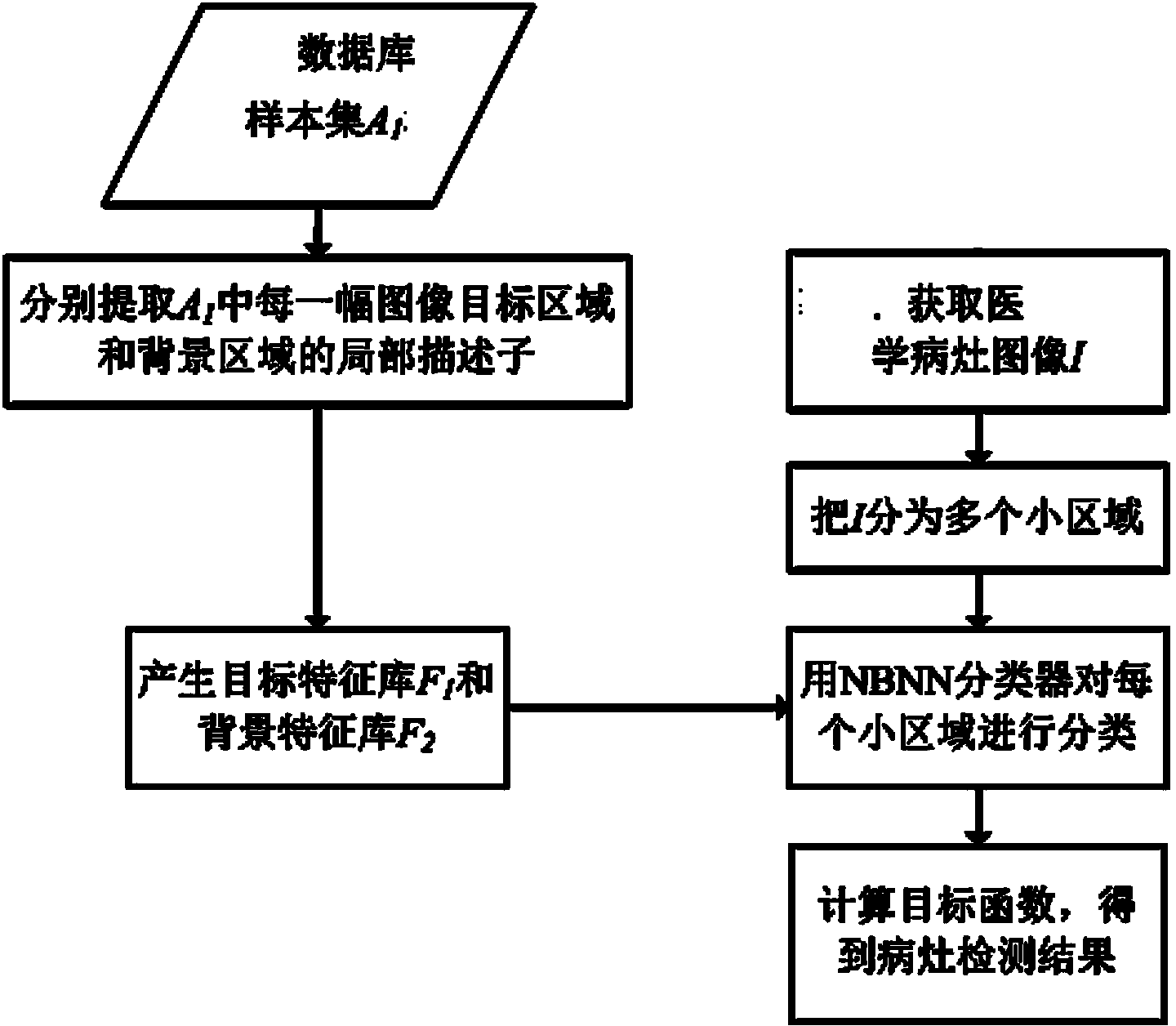
[0048] A kind of nonparametric automatic detection method of focal lesion of the present invention, such as figure 1 As shown, the sample set A is constructed in advance by using the medical lesion image that has been delineated as a sample. 1 Database, respectively extract sample set A 1 The local descriptors of the target area and the background area of ​​each image in the image, get the sample set A 1 The target feature library F composed of all target region local descriptors in 1 and sample set A 1 The background feature library F composed of local descriptors of all background regions in 2 .

[0049] Specifically, the sample set A is obtained as follows: 1 The target feature library F 1 and background feature library F 2 :

[0050] (0.1) Define the lesion area in the medical lesion image as the sample as the target area, and the area outside the lesion is the background area, and extract its local descriptor for each pixel in the target area; the same for each p...

Embodiment 2

[0085] The method of the present invention is described with a specific embodiment.

[0086] The database used in this embodiment has 458 liver CT image sample sets, including: 178 liver cancers, 98 liver cysts, and 182 hepatic hemangiomas, and each CT image has been manually delineated.


PUM


Abstract

Disclosed is a nonparametric automatic detection method of focal niduses. The method comprises the steps of using medical nidus images with the niduses marked as samples in advance, building a database of a sample set A1, extracting a local descriptor of the target region and the background region of each image in the sample set A1 respectively, and obtaining a target characteristic bank F1 composed of all the local descriptors of the target regions in the sample set A1, and a background characteristic bank F2 composed of all the local descriptors of the background regions in the sample set A1. A processing method comprises the following specific steps: (1) dividing the medical nidus images I to be processed into a plurality of sub-regions, (2) classifying each sub-region through an NBNN classifier, and (3) computing an objective function to obtain a detection result of the niduses. According to the nonparametric automatic detection method of the focal niduses, a parameterized model needs not to be built in advance, the local descriptors of the images need not to be quantified, the application is flexible, the resolution capability to the local descriptors of the images is strong, and the focal niduses can be accurately detected.

Description

technical field [0001] The invention relates to an automatic detection method for focal lesions in medical images, in particular to a non-parameter automatic detection method for focal lesions in medical images. Background technique [0002] In clinical diagnosis, lesion detection and segmentation in medical images is very important, because the extracted lesion area can provide doctors with the anatomical structure information of the lesion, and provide a basis for subsequent treatment and patient tracking. However, manually detecting and segmenting the lesion area is very time-consuming. Because some lesion tissue has no obvious contrast with the surrounding normal tissue, the lesion area segmented by different clinical experts may be different, which will easily increase the difficulty of subsequent diagnosis and tracking. [0003] In view of the shortage of manual detection and segmentation of lesion regions, researchers have proposed many methods for automatic detection...

Claims


Application Information

Patent Timeline
IPC IPC(8): G06K9/62A61B6/03
Inventor 阳维黄美燕冯前进佘广南卢振泰陈武凡
Owner SOUTHERN MEDICAL UNIVERSITY
Features
  • Generate Ideas
  • Intellectual Property
  • Life Sciences
  • Materials
  • Tech Scout
  • Unparalleled Data Quality
  • Higher Quality Content
  • 60% Fewer Hallucinations
Learn More