Blood perfusion separation detecting and imaging method for bone surface capillary

An imaging method and microvascular technology, applied in the fields of medical science, acoustic wave diagnosis, infrasound wave diagnosis, etc., can solve the problems of low spatial resolution, low CTR, bone surface guided wave order, complex frequency-variant aliasing, etc.

Active Publication Date: 2013-11-06
XI AN JIAOTONG UNIV
View PDF4 Cites 15 Cited by
  • Summary
  • Abstract
  • Description
  • Claims
  • Application Information

AI Technical Summary

Problems solved by technology

[0004] Although the rotation and movement of the ultrasound probe will inevitably cause oblique incidence imaging, which can provide clinically obtained multi-slice fine information that cannot be obtained by other imaging equipment, but at the same time the beam excites the bone surface will produce the unique problem of multi-order and multi-mode Lamb wave interference imaging. limit
Moreover, the frequency-thickness product corresponding to the central frequency of the clinical conventional ultrasound probe in the diagnosis of the musculoskeletal system is generally higher than that of simulation or isolated bone surface guided wave research, and the array probe excites bone surface guided wave orders and frequency-variant aliasing are more complicated.
At the same time, conventional contrast-enhanced ultrasound imaging methods still suffer from conventional problems such as low detection sensitivity of microbubbles, low spatial resolution, low CTR, low SNR of TIC, etc.

Method used


Image

  • Blood perfusion separation detecting and imaging method for bone surface capillary
  • Blood perfusion separation detecting and imaging method for bone surface capillary
  • Blood perfusion separation detecting and imaging method for bone surface capillary

Examples


Embodiment Construction

[0048] The present invention will be described in detail below in conjunction with the drawings.

[0049] The method for detecting and imaging the blood perfusion separation of bone surface capillaries includes the following steps:

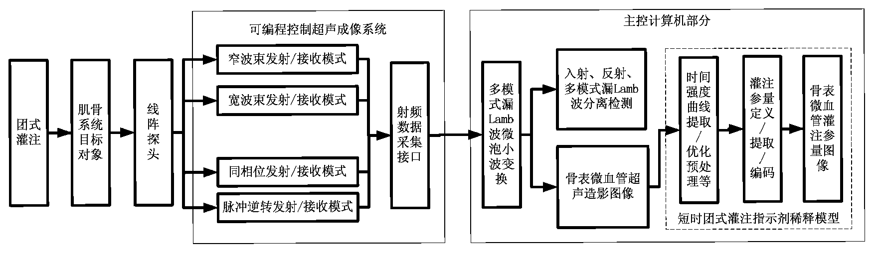
[0050] Step 1. On the programmable control ultrasound imaging system, according to the specific goals and clinical requirements of the musculoskeletal system, program and control the narrow / wide beam transmitting and receiving mode, and the same phase / pulse reverse phase setting mode;

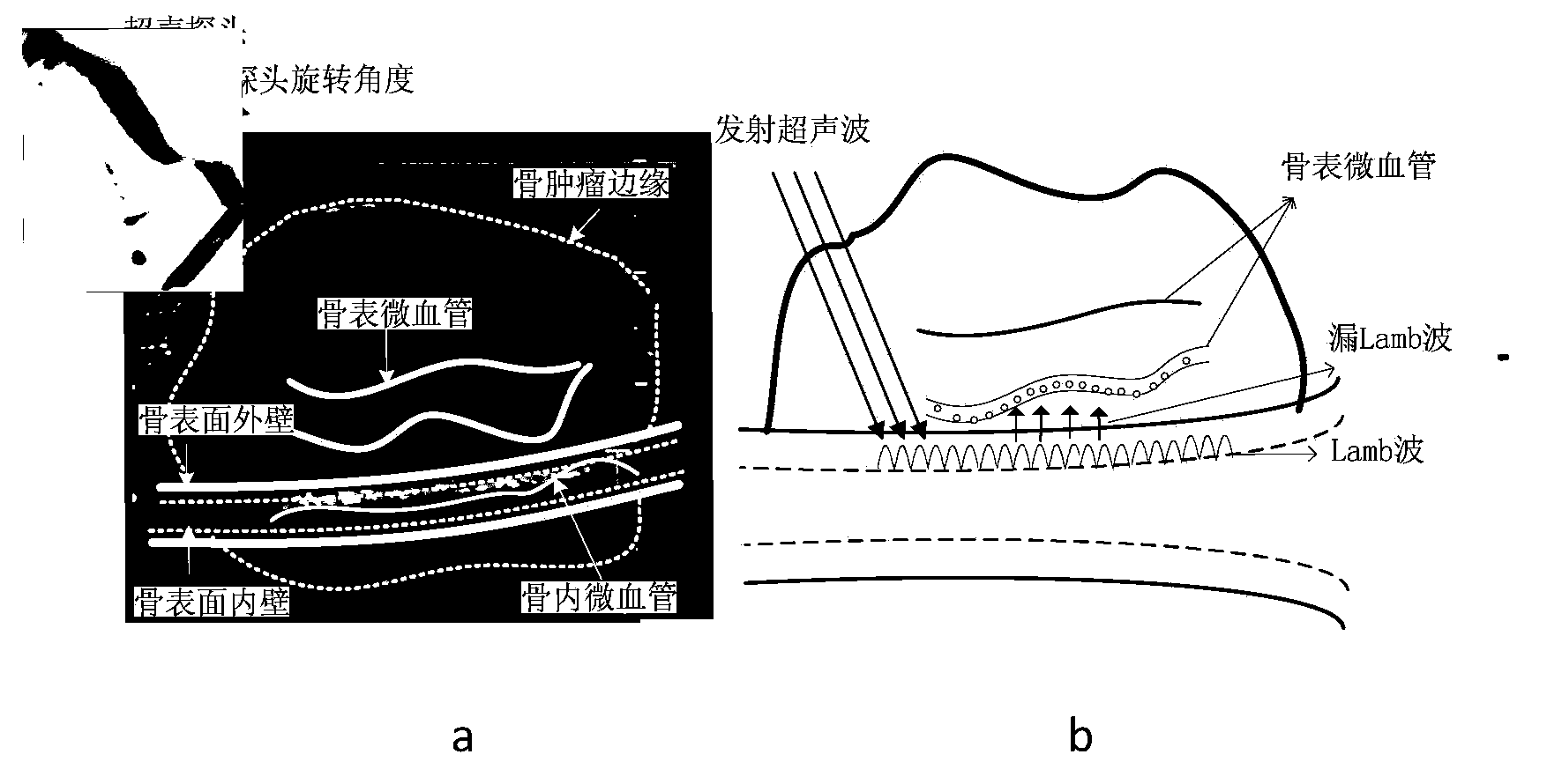
[0051] Step 2. On the main control computer platform, based on the Rayleigh-Lamb dispersion equation, namely formulas 1, 2 and the Morgen model modified Herring-Trilling microbubble vibration equations, namely formulas 3, 4, construct the bone surface multi-mode leaky Lamb wave microbubble Mother wavelet

[0052] tan(qh) / tan(ph)=(4k pq) / (q-k) (1, S P mode)

[0053] tan(qh) / tan(ph)=(q-k) / (4k pq) (2, A P mode)

[0054] ρ R · R · · + 3 2 ρ R ·...


PUM


Abstract

A blood perfusion separation detecting and imaging method for a bone surface capillary comprises the following steps: programming transmitting and receiving beams meeting the bone surface multimode leakage Lamb wave condition, constructing bone surface multimode leakage Lamb wave microbubble mother wavelets, then achieving mode separation detection of incidence, reflection and multi-mode leakage Lamb wave aliasing RF (Radio Frequency) echo signals by adopting the bone surface multimode leakage Lamb wave microbubble wavelet method, and finally achieving radiography imaging and blood perfusion parameter imaging of the transformation capillary for the bone surface multimode leakage Lamb wave microbubble wavelets. The blood perfusion separation detecting and imaging method can effectively improve the resolution and the radiography tissue ratio and suppress aliasing interference of the bone surface multimode leakage Lamb waves to radiography imaging of the bone surface tissue capillary, thereby achieving great significance on early diagnosis and evaluation of malignant bone tumors.

Description

Technical field [0001] The invention relates to the technical field of ultrasound imaging, in particular to a method for detecting and imaging the blood flow perfusion separation of the microvessels by the bone surface multi-mode leakage Lamb wave microbubble wavelet transform. Background technique [0002] Because the ultrasound imaging method has the advantages of non-radioactive, low price, good real-time performance, and good reproducibility, it has irreplaceable value and significance for the diagnosis and treatment of musculoskeletal system diseases, and provides reliable intervention basis for clinicians, especially for Major malignant diseases such as bone tumors. In view of the fact that many bone surfaces are non-planar, when the ultrasound probe moves and the angle is deflected, the bone surface is often accompanied by the propagation of multi-mode leaky Lamb waves. Due to many limitations, the current research on the dispersion and propagation characteristics of bone...

Claims


Application Information

Patent Timeline
Patent Type & Authority Applications(China)
IPC IPC(8): A61B8/00
Inventor 万明习王弟亚翟宇钟徽
Owner XI AN JIAOTONG UNIV
Why Eureka
Try Eureka