Curve fiber network structural morphology feature measurement method based on digital image processing

A technology of fiber network and shape characteristics, applied in the field of biological experiment image data analysis, can solve the problems that the shape characteristics of a single fiber cannot be obtained, and the shape information of the fiber network cannot be obtained.

Active Publication Date: 2014-03-26
裘钧
View PDF6 Cites 8 Cited by
  • Summary
  • Abstract
  • Description
  • Claims
  • Application Information

AI Technical Summary

Problems solved by technology

However, image processing methods based on tracking algorithms or SOACs algorithms can only extract single fibers, but cannot obtain the morphology information of the entire fiber network.
[0008] In addition, there are many other methods for processing curved fiber images, such as snake model-based algorithms to obtain smooth skeleton structures, and three-dimensional line structure enhancement filters to extract line structures from other structures. Morphological characteristics of fibers

Method used


Image

  • Curve fiber network structural morphology feature measurement method based on digital image processing
  • Curve fiber network structural morphology feature measurement method based on digital image processing
  • Curve fiber network structural morphology feature measurement method based on digital image processing

Examples


Embodiment 1

[0119] Analysis of images of real cellular stress fiber networks

[0120] The IFS algorithm of the present invention is applied to analyze real cell stress fiber network images. The skeleton network of actin filaments of osteoblasts (Osteoblast cell line: MC3T3) was labeled with XX fluorescence, and the microscopic images were obtained by observing under a laser confocal microscope, see Figure 5 (a). The center of the image is the center of the cell, and the periphery of the image is close to the edge of the cell. The size of each pixel in the image is 0.154 microns.

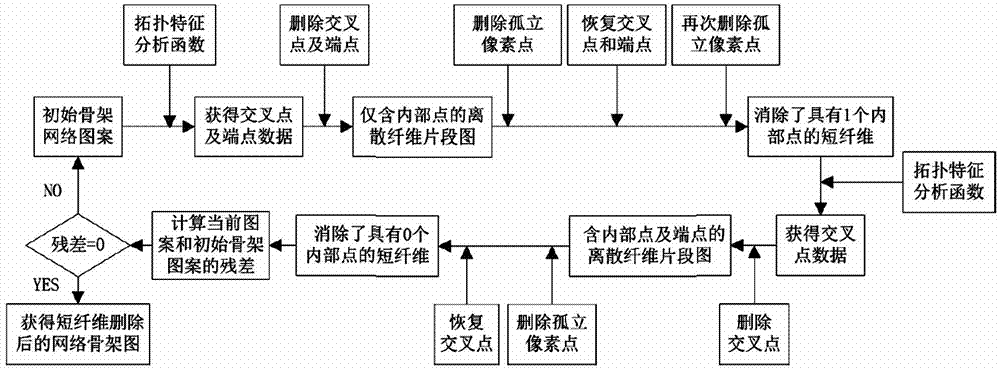
[0121] The process of analyzing the real cellular stress fiber skeleton network with the IFS program mainly includes:

[0122] Figure 5 (b) for image preprocessing;

[0123] Figure 5 (c) To delete weak out-of-plane fiber signals and skeletalization;

[0124] Figure 5 (d) Topological classification for skeleton pixels;

[0125] Figure 5 (e) to remove short fibers;

[0126] Figure 5 (f) Recognitio...


PUM


Abstract

A curve fiber network structural morphology feature measurement method based on digital image processing includes the steps: firstly, skeletonize fiber networks to obtain fiber network skeleton patterns; secondly, classifying topological features of the fiber network skeleton patterns and identifying single fibers and fiber crossing points in the fiber networks; thirdly, deleting short fibers and reserving network skeleton patterns of trunk fibers; fourthly, recombining fiber fragments, identifying crossing, branching and overlapping relationships in the fiber networks and recombining fiber network graphs. Curve fiber network structural morphology features of sub-cellular scales are obtained by the aid of the steps. The measurement method can be used for quantitatively analyzing curve fiber network structures of the sub-cellular scales, and can analyze quantitative topologies of the curve network structures in microscopic images without relying on human intervention and judgment after fiber structures in the curve networks are identified and segmented.

Description

technical field [0001] The invention belongs to the field of image data analysis of biological experiments, in particular to a digital image processing-based method for measuring the topography characteristics of curved fiber network structures applied at the subcellular scale. Background technique [0002] A large number of biological experiments have accumulated a large number of experimental pictures. In the past, biological research was mainly qualitative, and research conclusions were generally drawn through direct observation or simple quantitative analysis. The generation of more and more experimental images has brought us into an era of big data. Mining the quantitative laws behind the data has become a huge challenge for us. Data-driven research has brought biology to the quantitative era. With the vigorous development of medical image processing, the development of biological image processing does not seem to match the development of the field of biology. A type o...

Claims


Application Information

Patent Timeline
Patent Type & Authority Applications(China)
IPC IPC(8): G01B11/24
Inventor 裘钧李芳芳
Owner 裘钧
Why Eureka
Try Eureka