Eye fundus image characteristics extraction method for diabetic retinopathy

A diabetic retinal and fundus image technology, applied in image analysis, image data processing, instruments, etc., can solve problems such as uneven illumination and low blood vessel contrast

Inactive Publication Date: 2014-06-18
NANJING UNIV OF AERONAUTICS & ASTRONAUTICS +1
View PDF4 Cites 79 Cited by
  • Summary
  • Abstract
  • Description
  • Claims
  • Application Information

AI Technical Summary

Problems solved by technology

[0004] Aiming at the shortcomings of uneven illumination and low blood vessel contrast in fundus images at this stage, a method for processing fundus images of diabetic retinopathy is provided by using digital non-mydriatic fundus ...

Method used


Image

  • Eye fundus image characteristics extraction method for diabetic retinopathy
  • Eye fundus image characteristics extraction method for diabetic retinopathy
  • Eye fundus image characteristics extraction method for diabetic retinopathy

Examples


Embodiment 1

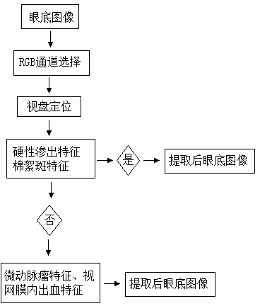
[0063] The fundus image used in this embodiment is taken by the non-mydriatic fundus camera TopconNW100, such as Figure 3a Shown. (1) RGB channel selection

[0064] The pigments contained in the various physiological structures of the fundus have different absorption characteristics, and the penetration performance of monochromatic light of different wavelengths in the fundus is also different. According to the characteristics of different DR lesions, they are divided into red image characteristics (MAs, Hs) and white images Features (EXs, CWs), for different detection targets, select the appropriate color space representation or sub-channel according to the results of the spectral feature analysis.

[0065] The optic disc has the highest visibility under 628nm red light. Under the light of this wavelength, the edges of the optic disc are clear, the blood vessels from the optic disc are poorly visible, the nerve fibers almost disappear, and the optic disc appears as a uniform ref...


PUM


Abstract

The invention discloses an eye fundus image characteristics extraction method for diabetic retinopathy by utilizing an eye fundus image shot by a digital mydriatic-free eye fundus camera. The method comprises the following steps of (1) selecting an RGB channel of the eye fundus image; (2) positioning the optic disk of the eye fundus image; (3) extracting the hard exudate characteristics and the cotton wool spot characteristics of the eye fundus image, generating an extracted eye fundus image if at least one characteristic is discovered, if no characteristic is discovered, extracting microaneurysm characteristics and retinal hemorrhage characteristics, and generating an extracted eye fundus image. The method belongs to a non-intrusive technology, and is simple, rapid, convenient and effective, and the processed image is clear and obvious in symptom, so the diagnosis of a doctor is facilitated.

Description

Technical field [0001] The invention relates to the field of digital image processing, in particular to a method for extracting fundus image features of diabetic retinopathy. Background technique [0002] Diabetic retinopathy (Diabetic Retinopathy, hereinafter referred to as DR) is the complication with the highest incidence in diabetic patients and has the greatest impact on vision. It is currently the main cause of new blindness in adults aged 20-65. Clinically, DR is divided into nonproliferative diabetic retinopathy (NPDR) (or simple or background type) and proliferative diabetic retinopathy (PDR) based on whether there is retinal neovascularization. In the non-proliferative phase, microaneurysms (hereinafter referred to as MAs), intraretinal hemorrhages (Haemorrhages, hereinafter referred to as Hs) and hard exudates (Hard exudates, hereinafter referred to as EXs) caused by capillary leakage will appear in the retina of patients. Cotton wool spots (CottonWoolSpots, hereinaft...

Claims


Application Information

Patent Timeline
IPC IPC(8): G06K9/62G06K9/46G06T7/40
Inventor 沈建新高玮玮庞杰周薇
Owner NANJING UNIV OF AERONAUTICS & ASTRONAUTICS
Why Eureka
Try Eureka