
Image guide method implemented by aid of two-dimensional images

An image-guided, three-dimensional image technology, applied in the field of image guidance, can solve problems such as difficult to find, deviation from the implant position, and increased patient risk and cost.

Inactive Publication Date: 2014-06-25
WEIDU MEDICAL SYST
View PDF0 Cites 7 Cited by
  • Summary
  • Abstract
  • Description
  • Claims
  • Application Information

AI Technical Summary

Problems solved by technology

However, the implantation of markers is an invasive clinical method, which will increase certain risks and costs for patients, and its scope of application is also limited
Clinically, about a week after the marker is implanted in the body, preparations for radiotherapy such as three-dimensional image acquisition are performed after the implant is settled, so there is a certain delay in the treatment.
And the implanted marker is prone to shift or fall off in the tissue, that is, to deviate from its implanted position
In this case, the use of these markers will cause large positioning errors and are not easy to be found

Method used


Image

  • Image guide method implemented by aid of two-dimensional images
  • Image guide method implemented by aid of two-dimensional images
  • Image guide method implemented by aid of two-dimensional images

Examples


Embodiment Construction

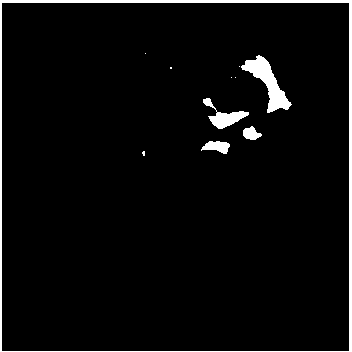
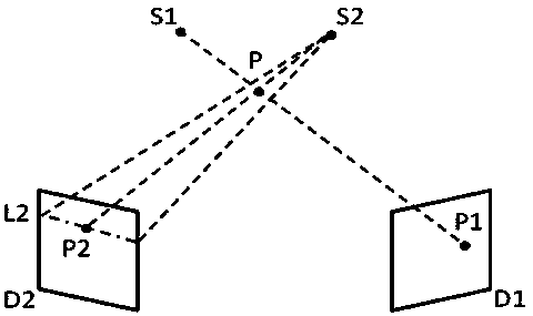
[0029] In this method, the location deviation is determined by searching some real-time feature areas in the real-time map, and comparing the positions of the real-time feature areas in the real-time map with the reference feature areas in the DRR. These reference feature areas are small areas in 3D images, and their projections in 2D images (including real-time images and 2D DRR) are small areas with characteristics different from surrounding areas. The specific feature description depends on the image mode and image comparison (also known as registration or fusion, etc.) algorithm, such as in X-ray two-dimensional images, the reference feature area can have a large and unique change in grayscale small areas, such as figure 1 The two regions shown in . The reference feature area selected here is the reference feature area of ​​a non-artificially implanted marker. Image guidance by implanting metal or other markers in radiotherapy has been reported for a long time and has b...


PUM


Abstract

The invention discloses an image guide method implemented by the aid of two-dimensional images. The image guide method includes steps of selecting and screening reference feature regions; searching real-time feature regions; performing positioning and computing on the basis of the reference feature regions and the real-time feature regions; estimating deformation and errors, and the like. The image guide method has the advantages that two-dimensional DRRs (digitally reconstructed radiographs) need to be generated according to three-dimensional images of lesions of patients when image guide is carried out by the aid of two-dimensional images, and then the feature regions in real-time images which are acquired in real time are compared to feature regions in the two-dimensional DRRs, so that deviation among current locations and preset locations can be determined; markers do not need to be implanted in the patients when the patients are examined by the aid of the image guide method, accordingly, suffering can be relieved for the patients, and treatment risks and the treatment cost can be reduced for the patients.

Description

technical field [0001] The invention relates to the field of image guidance, in particular to an image guidance method using two-dimensional images. Background technique [0002] The basic purpose of image guidance is to reproduce a preset patient position during actual treatment or treatment, referred to as preset position. Taking radiotherapy as an example, doctors collect three-dimensional CT images of patients, formulate radiotherapy plans based on the images, and select a point in the images as the treatment center, which is called the isocenter. During treatment, the anatomical positions represented by these centers need to be placed at the center of the treatment equipment, usually the medical electron linear accelerator (also known as the isocenter, the isocenter of the equipment). The preset body position refers to the patient's body position when the anatomical position represented by the isocenter set by the radiotherapy plan is located at the isocenter of the eq...

Claims


Application Information

Patent Timeline
IPC IPC(8): A61B6/03
Inventor 母治平
Owner WEIDU MEDICAL SYST
Learn More