Method for extracting and analyzing nidus areas in pneumoconiosis gross imaging

An analysis method, pneumoconiosis technology, applied in the direction of instruments, character and pattern recognition, computer components, etc., can solve the problems of cumbersome operation, low efficiency, large measurement error, etc., and achieve the effect of high efficiency

Active Publication Date: 2014-06-25
WUHAN TIANREN IMAGE TECH CO LTD
View PDF3 Cites 16 Cited by
  • Summary
  • Abstract
  • Description
  • Claims
  • Application Information

AI Technical Summary

Problems solved by technology

At present, the analysis of gross pathological images relies entirely on manual operatio

Method used


Image

  • Method for extracting and analyzing nidus areas in pneumoconiosis gross imaging
  • Method for extracting and analyzing nidus areas in pneumoconiosis gross imaging
  • Method for extracting and analyzing nidus areas in pneumoconiosis gross imaging

Examples


Embodiment 1

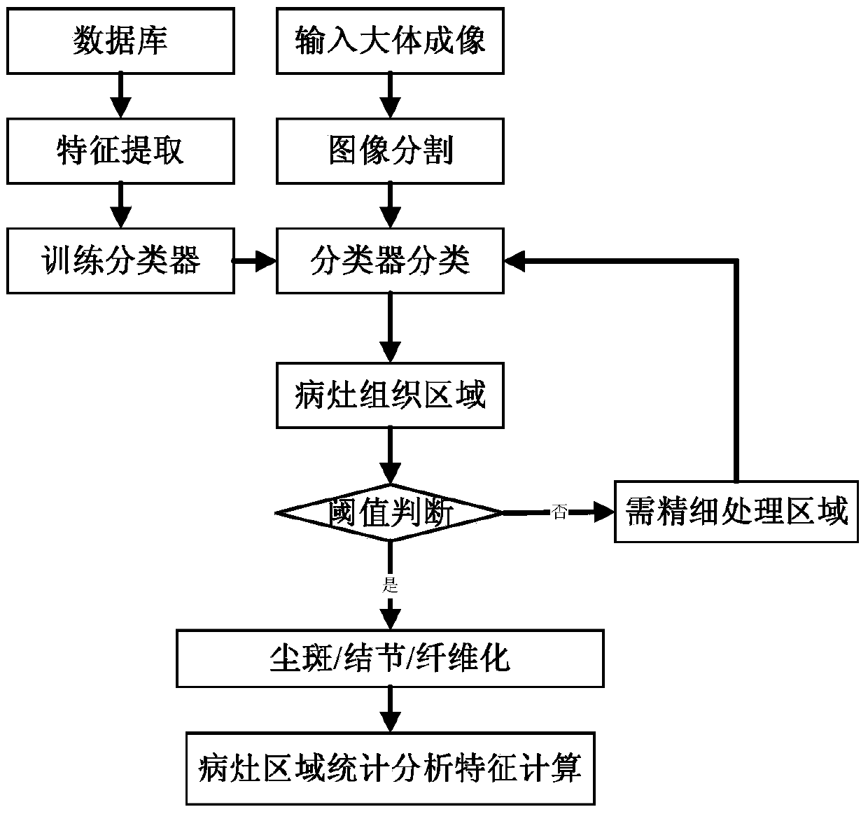
[0033] The extraction and analysis method of the lesion area in the pneumoconiosis gross imaging, the steps are as follows:

[0034] S1. Extract features:

[0035] S11. connect the pneumoconiosis gross imaging database with the computer,

[0036] S12. Calculate the center coordinates (c x ,c y ), taking the center of the region as the starting point, draw 36 rays evenly every 10°, and intersect with the corresponding points on the boundary of the region respectively to obtain 36 line segments, divide each line segment into 5 equal parts, divide each line segment into The bisection points of each are connected respectively, and the area is divided into 5 ring parts, and the average gray value of each ring part is calculated to form a 5-dimensional feature vector, and the difference operation is performed on the 5-dimensional feature vector to obtain a 4-dimensional feature vector, and the 5-dimensional feature vector is obtained The dimensional feature vector and the 4-dimen...

Embodiment 2

[0065] The extraction and analysis method of the lesion area in the pneumoconiosis gross imaging, the steps are as follows:

[0066] S1. Extract features:

[0067] S11. connect the pneumoconiosis gross imaging database with the computer,

[0068] S12. Calculate the center coordinates (c x ,c y ), taking the center of the region as the starting point, draw 36 rays evenly every 10°, and intersect with the corresponding points on the boundary of the region respectively to obtain 36 line segments, divide each line segment into 5 equal parts, divide each line segment into The bisection points of each are connected respectively, and the area is divided into 5 ring parts, and the average gray value of each ring part is calculated to form a 5-dimensional feature vector, and the difference operation is performed on the 5-dimensional feature vector to obtain a 4-dimensional feature vector, and the 5-dimensional feature vector is obtained The dimensional feature vector and the 4-dimen...


PUM


Abstract

The invention relates to a method for extracting and analyzing nidus areas in pneumoconiosis gross imaging. The method comprises the following steps that the feature vector F1 of a 2k-1 dimension is calculated, pre-training is carried out, a feature vector F2 is calculated, a support vector machine classifier h0 used for judging whether an area is the nidus areas or not is trained, multi-classification support vector machine classifiers h1-hk used for judging an area class are trained, the pneumoconiosis gross imaging is input, the features of the nidus areas are extracted, the nidus areas are classified, and statistical characteristic calculation is carried out on the nidus areas. Spatial expansion is carried out on image pixels to from small surface element areas, the surface element areas are classified by the trained classifiers, the surface element areas with the low classification confidence coefficient are finely processed, and the different nidus areas are extracted accurately. The method is high in efficiency, small in error and free of man-machine interaction.

Description

technical field [0001] The invention relates to a method for extracting and analyzing regions in images, in particular to a method for extracting and analyzing lesion regions in gross imaging of pneumoconiosis. Background technique [0002] Pneumoconiosis (pneumoconiosis) is a systemic disease mainly caused by diffuse fibrosis of lung tissue caused by long-term inhalation of productive dust in occupational activities and retention in the lungs. At present, the number of pneumoconiosis patients in my country is large and the growth rate is fast. Ranked first in the world. [0003] At present, the diagnosis of pneumoconiosis mainly includes X-ray transmission imaging (chest radiography) diagnosis and pathological diagnosis. Although chest X-ray diagnosis is a non-destructive diagnostic method, due to the reason of the imaging method, the interpretation results have large deviations, so it is only suitable for the screening of pneumoconiosis. The final diagnosis of pneumoconios...

Claims


Application Information

Patent Timeline
IPC IPC(8): G06K9/46G06K9/62
Inventor 明德烈
Owner WUHAN TIANREN IMAGE TECH CO LTD
Why Eureka
Try Eureka