Method for establishing image data through nuclear magnetic resonance

A technology of image data and method establishment, applied in the field of medical imaging, can solve problems such as low accuracy and repeatability of ultrasound, untimely isotope, quantitative response data, etc., and achieve the effect of avoiding waste

Inactive Publication Date: 2014-07-16
SHANGHAI SIXTH PEOPLES HOSPITAL
View PDF1 Cites 6 Cited by
  • Summary
  • Abstract
  • Description
  • Claims
  • Application Information

AI Technical Summary

Problems solved by technology

[0003] In order to solve the problem that the current stress ultrasound and isotopes have low ultrasound accuracy and repeatability when obtaining myocardial perfusion data, and isotopes cannot respond to data in a timely and quantitative manner, a method for establishing image data using nuclear magnetic resonance is now provided.

Method used


Image

  • Method for establishing image data through nuclear magnetic resonance
  • Method for establishing image data through nuclear magnetic resonance

Examples


Embodiment Construction

[0020] In order to make the object, technical solution and advantages of the present invention clearer, the implementation manner of the present invention will be further described in detail below in conjunction with the accompanying drawings.

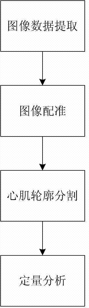
[0021] An embodiment of the present invention provides a method for establishing image data using nuclear magnetic resonance, including the following steps,

[0022] Step 1, extracting the nuclear magnetic resonance image data of the target area when the contrast agent passes first.

[0023] Step 2, registering the image sequence to eliminate the displacement deviation of the target area in the image sequence. The registration of the image sequence includes calculating the registration template image corresponding to each image in the image sequence through a non-rigid registration algorithm based on pseudo-real information, and using the Markov random field model to compare the target image with the corresponding Image registration i...


PUM


Abstract

The invention discloses a method for establishing image data through nuclear magnetic resonance and belongs to the technical field of medical images. The method comprises the following steps that nuclear magnetic resonance image sequential data of a target zone in the first pass process of a contrast agent are extracted; registration is conducted on an image sequence, so that displacement deviation of the target zone in the image sequence is eliminated; the myocardium outline in the target zone of an image is segmented, so that an inner outline and an outer outline of the myocardium are obtained; quantitative analysis is conducted, the myocardium in the image is segmented into a plurality of partial zones according to an ultrasonic cardiogram seventeen-segmentation division method along the outline, a pixel mean value of each zone is calculated, and a data curve of the relation of the signal intensity and time is obtained. The nuclear magnetic resonance image data are collected, image registration is conducted, the myocardium outline image data are obtained through segmentation, quantitative analysis is conducted, a data model and an accurate data analysis platform are established, and therefore waste of resources is avoided.

Description

technical field [0001] The invention relates to the technical field of medical imaging, in particular to a method for establishing image data using nuclear magnetic resonance. Background technique [0002] Stress ultrasound and isotopes are a common way to obtain myocardial perfusion data. However, ultrasound is highly subjective, with low accuracy and repeatability; isotopes cannot always and quantitatively reflect the data information of myocardial perfusion and metabolic status. Contents of the invention [0003] In order to solve the problem that the current stress ultrasound and isotopes have low ultrasound accuracy and repeatability when obtaining myocardial perfusion data, and isotopes cannot respond to data in a timely and quantitative manner, a method for establishing image data using nuclear magnetic resonance is now provided. The specific technical scheme is as follows: [0004] A method for establishing image data using nuclear magnetic resonance, comprising ...

Claims


Application Information

Patent Timeline
Patent Type & Authority Applications(China)
IPC IPC(8): G06T7/00
Inventor 魏盟杨新潘静薇邵叶秦王健
Owner SHANGHAI SIXTH PEOPLES HOSPITAL
Why Eureka
Try Eureka