X-ray light field imaging and calibrating method based on pinhole array

A pinhole array and light field imaging technology, which is applied in the fields of radiological diagnosis instruments, medical science, diagnosis, etc., can solve the problems of overlapping images of different depths, loss of depth information in X-ray images, and indistinguishability.

Inactive Publication Date: 2014-09-03
ZHEJIANG GONGSHANG UNIVERSITY
View PDF2 Cites 12 Cited by
  • Summary
  • Abstract
  • Description
  • Claims
  • Application Information

AI Technical Summary

Problems solved by technology

[0006] Aiming at the deficiencies of the prior art, the present invention extends the light field imaging theory of visible light to X-ray imaging to solve the problem that the depth information of X-ray images is lost in clinical diagnosis and treatment, resulting in overlapping and indistinguishable images of different depths, and proposes a X-ray light field imaging and calibration method based on pinhole array without loss of depth information, no image overlap, low dose, fast imaging, and meeting the imaging quality requirements of X-ray images in clinical diagnosis and treatment

Method used


Image

  • X-ray light field imaging and calibrating method based on pinhole array
  • X-ray light field imaging and calibrating method based on pinhole array
  • X-ray light field imaging and calibrating method based on pinhole array

Examples


Embodiment 1

[0046] An X-ray light field imaging and calibration method based on a pinhole array, comprising the following steps:

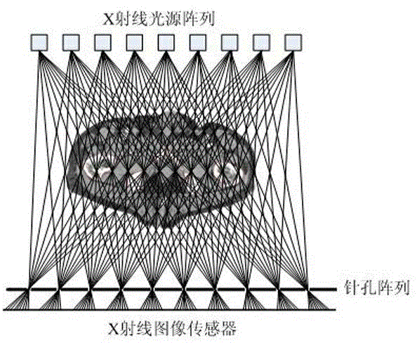
[0047] Step 1: Establish an X-ray light field imaging model based on a pinhole array, the schematic diagram of the imaging plane structure is as follows figure 1 As shown, it includes an X-ray light source array, a pinhole array and an X-ray image sensor. The X-ray image sensor adopts a digital image sensor that can directly image X-rays. The X-ray image sensor array is a group of digital image sensors that can be directly imaged by X-rays. In this embodiment, what the image sensor adopts is a CCD or CMOS chip.

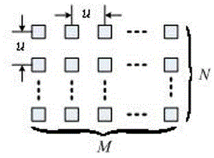
[0048] The X-ray light source array is a two-dimensional planar array of M×N orthogonal arrangements composed of multiple X-ray light sources, such as figure 2 shown. The size of the X-ray light source array determines the angular resolution of the X-ray light field imaging.

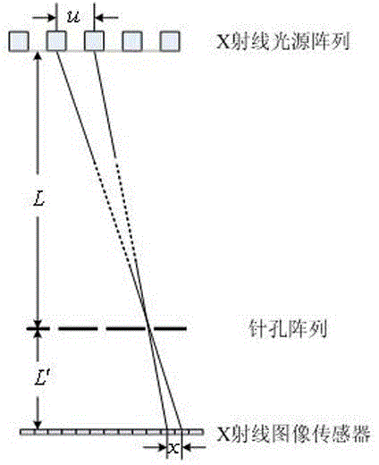
[0049] according to image 3 In the X-ray pinhole imaging geometry, ...


PUM


Abstract

The invention discloses an X-ray light field imaging and calibrating method based on a pinhole array. The X-ray light field imaging and calibrating method based on the pinhole array comprises the steps that (1) An X-ray light field imaging model based on the pinhole array is established and comprises an X-ray light source array, the pinhole array and an X-ray image sensor; (2) a linear exposure mapping image is established so that exposure of images of an X-ray light field can be normalized; (3) the pinhole center of each X-ray light field image is calibrated so that the central position of each pinhole image is extracted; (4) according to the center, obtained in the step (3), of each pinhole image, each X-ray light source image is extracted; (5) according to each X-ray light source image extracted in the step (4), the pixels at the same position in the pinhole images are extracted and reorganized into a frame of X-ray light source image; (6) the extracted X-ray light source images are converted into X-ray light source images which are orthogonally arranged. According to the X-ray light field imaging and calibrating method based on the pinhole array, the light field imaging principle of visible light is applied to X-ray imaging, and the requirement for the imaging quality of the X-ray images during clinical diagnosis and treatment is met.

Description

technical field [0001] The invention relates to light field imaging technology in the field of X-ray medical imaging, in particular to an X-ray light field imaging and calibration method based on a pinhole array. Background technique [0002] Light field imaging is based on traditional imaging equipment, through camera arrays, coded apertures or microlens arrays to achieve light field acquisition. In terms of light field imaging based on camera arrays, M. Levoy of Stanford University built a 4D light field acquisition platform consisting of 128 cameras in 1996. [1] . In 2000, P.Debevec of the University of Southern California realized the 4D static variable light acquisition system for face acquisition for the first time. [2] , Professor Dai Qionghai of Tsinghua University in China built the first domestic variable light dynamic light field acquisition system with a diameter of 6 meters, including 40 cameras and 310 LED light sources [3] . In 2012, the project team and T...

Claims


Application Information

Patent Timeline
IPC IPC(8): A61B6/00
Inventor 林丽莉周文晖王秀萍
Owner ZHEJIANG GONGSHANG UNIVERSITY
Why Eureka
Try Eureka