Colon image segmenting method, and colon image segmenting device
An image segmentation and colon technology, applied in the field of medical image processing, can solve problems such as loss of intestinal breakage, and achieve the effect of improving integrity
- Summary
- Abstract
- Description
- Claims
- Application Information
AI Technical Summary
Problems solved by technology
Method used
Image

Examples
Embodiment 1
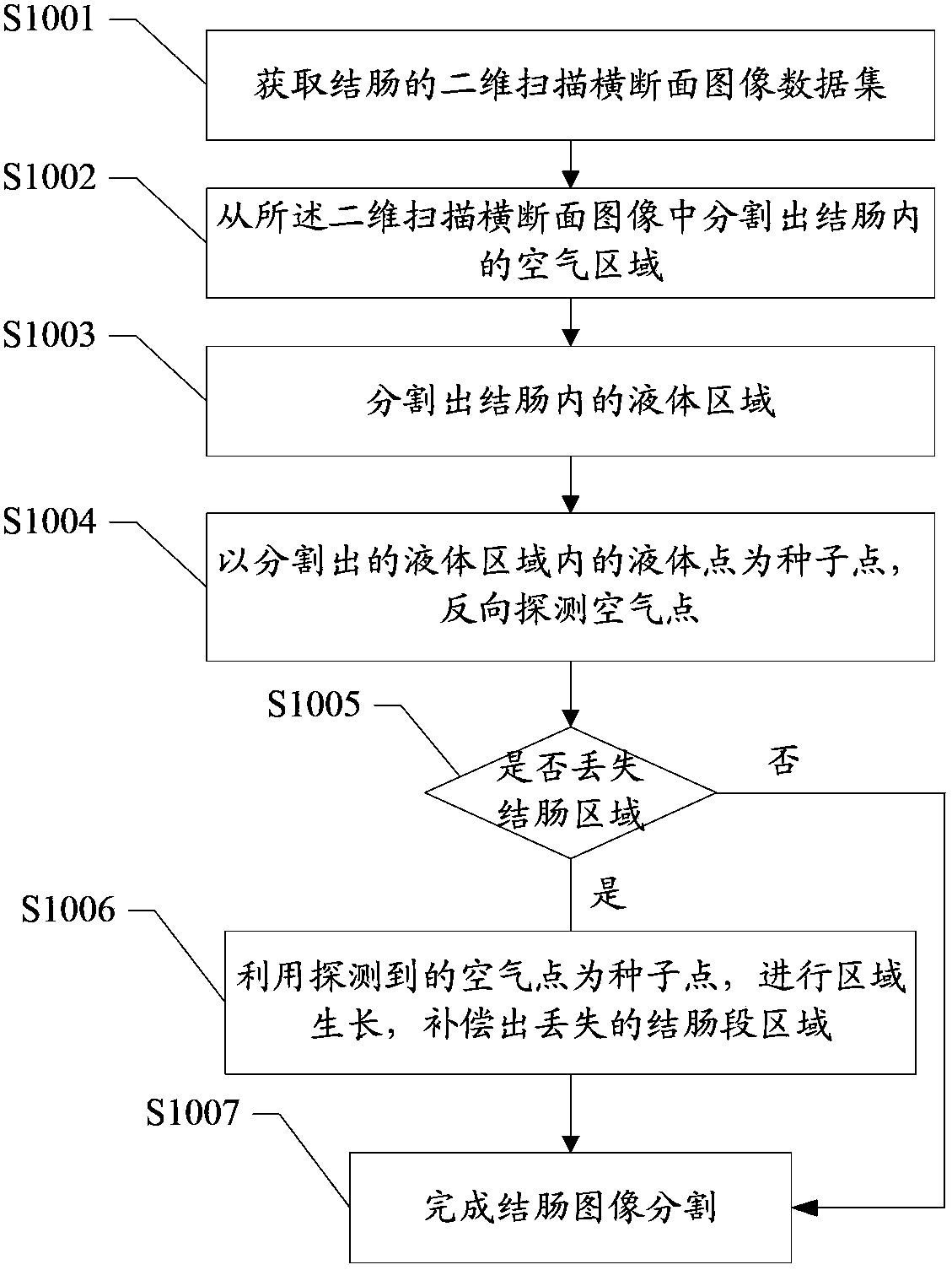
[0037] After segmenting the liquid area in the colon, using the liquid point in the segmented liquid area as the seed point, the air point is detected in the reverse direction to determine whether the colon segment area is lost, and when determining the missing colon segment area, use the detected The air point is the seed point for regional growth to compensate for the lost colon segment. The following reference figure 1 , To explain in detail through specific steps:
[0038] S1001: Obtain a two-dimensional scan cross-sectional image data set of the colon.
[0039] In a specific implementation, a variety of medical scanning equipment can be used to scan the abdomen of the subject to obtain a two-dimensional scanned cross-sectional image of the colon. In this embodiment, CT scan images are used.
[0040] S1002: Segment the air area in the colon from the two-dimensional scanned cross-sectional image.
[0041] In the specific implementation, the air area in the background can be remov...
Embodiment 2
[0053] The scope of colon examination usually includes the colon tissue and the rectum connected to the colon, where the lower end of the rectum is connected to the anus, and the upper end of the rectum is connected to the sigmoid colon at the lower end of the colon. The inventor found that due to the narrowness of the sigmoid colon, the existing segmentation of the colon image easily causes the rectal region located at the anus to be lost. In order to avoid the loss of the rectal region, in this embodiment, the human anatomy is used to determine whether the rectum is lost in the image after threshold segmentation, and when it is determined that the rectal region is lost, the rectal region is compensated.
[0054] Reference image 3 As shown in the flowchart of the colon image segmentation method, this embodiment specifically includes the following steps:
[0055] S3001: Obtain a two-dimensional scan cross-sectional image data set of the colon.
[0056] In specific implementations, ...
Embodiment 3
[0098] Reference Picture 10 In the colon image segmentation device shown, the colon image segmentation device 1000 includes: an image data acquisition unit 1001, a first segmentation unit 1002, a second segmentation unit 1003, and a first compensation unit 1004, wherein:
[0099] The image data acquisition unit 1001 is used to acquire a two-dimensional scanned cross-sectional image data set of the colon;
[0100] The first segmentation unit 1002 is configured to segment the air area in the colon from the two-dimensional scanned cross-sectional image;
[0101] The second dividing unit 1003 further divides the liquid area after the first dividing unit divides the air area in the colon area;
[0102] The first compensation unit 1004 is used to reversely detect air points with the liquid points of the segmented liquid area to determine whether the colon segment area is lost, and when the colon segment area is lost, use the detected air points as the seed point to perform the area Growing...
PUM
 Abstract
Description
Claims
Application Information
 - Generate Ideas
- Intellectual Property
- Life Sciences
- Materials
- Tech Scout
- Unparalleled Data Quality
- Higher Quality Content
- 60% Fewer Hallucinations
Browse by: Latest US Patents, China's latest patents, Technical Efficacy Thesaurus, Application Domain, Technology Topic, Popular Technical Reports.
© 2025 PatSnap. All rights reserved.Legal|Privacy policy|Modern Slavery Act Transparency Statement|Sitemap|About US| Contact US: help@patsnap.com