Eureka AIR delivers breakthrough ideas for toughest innovation challenges, trusted by R&D personnel around the world.

Calcification location method based on dual-view mammography

A dual-view, mammary gland technology, which is applied in the fields of radiological diagnosis instruments, image data processing, medical science, etc., can solve the problems of large patient trauma, limited promotion and popularization, lack of convenience and reliability, and achieves convenient and accurate operation and positioning. Accurate, fast and reliable technical support

Inactive Publication Date: 2017-04-26
湖北省肿瘤医院
View PDF3 Cites 0 Cited by
  • Summary
  • Abstract
  • Description
  • Claims
  • Application Information

AI Technical Summary

Problems solved by technology

[0003] At present, the full digital mammography system and the three-dimensional positioning biopsy system can be used to locate the preoperative hook wire of the calcification, and the resection of the calcification and the histopathological examination under the guidance of the positioning line are an effective and safe diagnostic method , but its expensive equipment investment and its innovative positioning method limit its promotion and popularization in my country
[0004] Due to the lack of convenient and reliable devices for the localization of tiny calcifications in the breast with negative palpation, large tissue excision biopsy or 1 / 4 mastectomy is often used clinically to remove calcifications, which has a certain degree of blindness and is harmful to patients. more traumatic

Method used


Image

  • Calcification location method based on dual-view mammography
  • Calcification location method based on dual-view mammography
  • Calcification location method based on dual-view mammography

Examples


Embodiment Construction

[0028] The present invention will be further described below in conjunction with specific examples and accompanying drawings.

[0029] This embodiment provides a method for locating calcifications based on dual-view mammography, which includes the following steps:

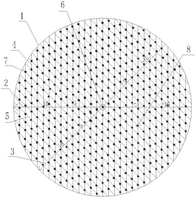
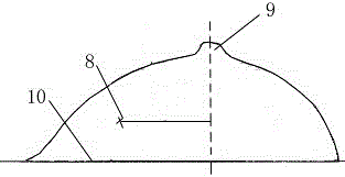
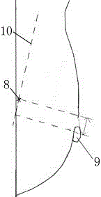
[0030] 1) Establish a coordinate system, the horizontal direction is the abscissa, and the oblique downward direction is the oblique coordinate. The angle between the oblique coordinate and the abscissa is consistent with the shooting angle of the MLO X-ray.

[0031] In order to be more in line with the doctor's habit of viewing films, a coordinate system is established for the left breast and the right breast respectively:

[0032] left breast: eg figure 1 As shown, the coordinate system 1 is composed of abscissa 2 and oblique coordinate 3. The abscissa 2 is the horizontal direction and the horizontal direction to the left is the positive direction. The included angle ranges from 35 degrees to 55 degrees, and th...


PUM


Abstract

The invention provides a two-viewing-angle mammography based calcification positioning method. The method includes the steps: 1) establishing a coordinate system with the horizontal direction serving as an x-coordinate and the obliquely downward direction serving as an oblique coordinate, wherein an included angle between the oblique coordinate and the x-coordinate is consistent with an MLO (mediolateral oblique)-position X-ray film photographing angle; 2) obtaining an absolute value of the x-coordinate according to a CC (craniocaudal)-position X-ray film, and obtaining an absolute value of the oblique coordinate according to an MLO-position X-ray film; 3) selecting the left breast or the right breast and respectively judging plus or minus of the x-coordinate and the oblique coordinate; 4) obtaining a coordinate of a calcification position by combining the step 2) with the step 3), and finding out a corresponding coordinate point in the coordinate system. The two-viewing-angle mammography based calcification positioning method has the advantages that the x-coordinate value and the oblique coordinate value are obtained by the aid of the CC-position X-ray film and the MLO-position X-ray film, the corresponding point is found out in the established coordinate system, the breast position corresponding to the corresponding point is the calcification position, and thereby operation is convenient and accurate, and the method provides reliable technical support for routine surgical biopsy.

Description

technical field [0001] The invention belongs to the field of calcification focus positioning, in particular to a method for calcification focus positioning based on dual-view mammography. Background technique [0002] In recent years, with the continuous improvement of breast imaging equipment, especially the update and improvement of the full digital mammography system, the imaging rate of small calcifications in the breast with negative clinical palpation has gradually increased. How to combine the images of tiny calcifications on X-ray films to perform localized biopsy of tiny calcifications in the breast with negative clinical palpation is the key to improving the early diagnosis rate and accurate resection rate of breast cancer without mass. [0003] At present, the full digital mammography system and the three-dimensional positioning biopsy system can be used to locate the preoperative hook wire of the calcification, and the resection of the calcification and the histo...

Claims


Application Information

Patent Timeline
Patent Type & Authority Patents(China)
IPC IPC(8): G06T7/00A61B6/00
Inventor 金立亭龚益平
Owner 湖北省肿瘤医院
Why Eureka
Eureka Blog
Learn More