Method and device for identifying organ blood vessels

A recognition method and blood vessel technology, applied in the field of medical image processing, can solve the problems of occupying large memory and video memory space, a large amount of calculation, and low efficiency of blood vessel recognition, and achieve the effects of reducing the amount of calculation, eliminating interference, and saving storage space

Active Publication Date: 2014-12-24
QINGDAO HISENSE MEDICAL EQUIP
View PDF4 Cites 4 Cited by
  • Summary
  • Abstract
  • Description
  • Claims
  • Application Information

AI Technical Summary

Problems solved by technology

[0003] The purpose of the embodiment of the present invention is to provide a method and device for organ blood vessel recognition to solve the following problems in the prior art of organ blood vessel recognition: when performing organ blood vessel recognition, the blood vessel recognition efficiency is low due to the large amount of calculation, and a large amount of time is occupied. Memory and video memory space

Method used


Image

  • Method and device for identifying organ blood vessels
  • Method and device for identifying organ blood vessels
  • Method and device for identifying organ blood vessels

Examples


Embodiment Construction

[0054] A method and device for identifying blood vessels in organs provided by the present invention will be described in more detail below with reference to the accompanying drawings and embodiments.

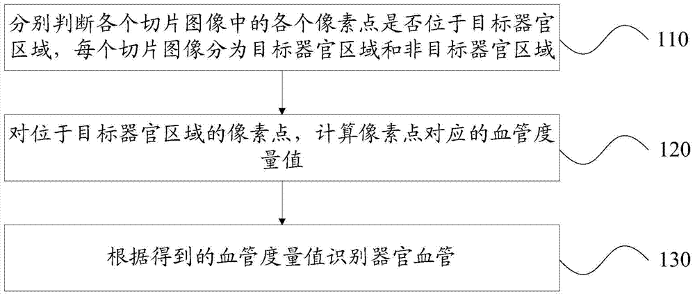
[0055] An embodiment of the present invention provides a method for identifying blood vessels in organs, such as figure 1 As shown, the specific steps are as follows:

[0056] Step 110: Determine whether each pixel point in each slice image is located in the target organ area, and each slice image is divided into a target organ area and a non-target organ area.

[0057] Wherein, the slice image may be a grayscale image, and correspondingly, the pixel value is represented by a grayscale value.

[0058] Wherein, the target organ refers to the organ to be identified for blood vessels.

[0059] Step 120: For the pixel points located in the target organ region, calculate the blood vessel metric values ​​corresponding to the pixel points.

[0060] Step 130: Identify organ vessels ...


PUM


Abstract

An embodiment of the invention discloses a method and a device for identifying organ blood vessels. The method includes respectively judging whether or not each pixel point in each section image is positioned in a target organ region, wherein each section image is divided into the target organ region and a non-target organ region; calculating blood vessel metrics corresponding to the pixel points positioned in the target organ regions; identifying the organ blood vessels according to the acquired blood vessel metrics. Whether or not the pixel points in the section images are positioned in the target organ regions is respectively judged, each section image is divided into the target organ region and the non-target organ region, only the blood vessel metrics corresponding to the pixel points positioned in the target organ regions are calculated, calculation of blood vessel metrics corresponding to all the pixel points is not needed, and the amount of calculation is reduced during calculation of the blood vessel metrics, so that processing efficiency of blood vessel identification is improved, data in need of storage is reduced during calculation, and storage space is saved. The problems of low organ blood vessel identification efficiency and occupation of substantial internal storage and video memory space are solved.

Description

technical field [0001] The invention relates to the field of medical image processing, in particular to a method and device for identifying blood vessels in organs. Background technique [0002] The body organ information obtained by computer tomography (Computer Tomography, CT) angiography is only a two-dimensional sliced ​​image. Usually, slice images need to be processed by an image processor (Graphic Processing Unit, GPU) to obtain a three-dimensional image, which shows the blood vessel structure in the organ. When the GPU processes the sliced ​​images, the computer central processing unit (Central Processing Unit, CPU) first reads all the sliced ​​images into the memory, and then transfers the sliced ​​images to be processed to the video memory. Usually, the GPU can use the Hessian matrix corresponding to the pixels and the eigenvalues ​​of the Hessian matrix to identify the vascular structure in the organ. Therefore, the GPU first needs to calculate the Hessian matri...

Claims


Application Information

Patent Timeline
Patent Type & Authority Applications(China)
IPC IPC(8): G06K9/00
Inventor 宋沂鹏杨杰陈永健
Owner QINGDAO HISENSE MEDICAL EQUIP
Why Eureka
Try Eureka