
Liver Segmentation Method in CT Image Based on Adaptive Surface Deformation Model

A surface deformation and CT image technology, applied in the image scanning field of liver segmentation, can solve the problems of partial balance of the model, overflow of the deformed model, inability to effectively segment the liver tissue, etc., and achieve the effect of ensuring smoothness and accuracy

Active Publication Date: 2017-07-14
ARIEMEDI MEDICAL SCI BEIJING CO LTD
View PDF3 Cites 0 Cited by
  • Summary
  • Abstract
  • Description
  • Claims
  • Application Information

AI Technical Summary

Problems solved by technology

[0004] 1. The initial position of the deformable model seriously affects the final segmentation result. If the model initialization is too small, it is easy to cause the model to reach local equilibrium or shrink to a point.
At this time, the liver tissue cannot be effectively segmented
[0005] 2. If the model initialization is too large, it is easy to cause the deformation model to overflow the segmentation area, resulting in over-segmentation
[0006] 3. Due to the influence of the smooth force of the deformable model, it cannot effectively enter the slender and sharp corners of the liver, which seriously affects the segmentation accuracy

Method used


Image

  • Liver Segmentation Method in CT Image Based on Adaptive Surface Deformation Model
  • Liver Segmentation Method in CT Image Based on Adaptive Surface Deformation Model
  • Liver Segmentation Method in CT Image Based on Adaptive Surface Deformation Model

Examples


Embodiment Construction

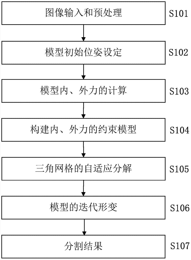
[0024] as attached figure 1 As shown, the flowchart of the present invention specifically includes the following steps:

[0025] Step S101, image preprocessing.

[0026] The original image is preprocessed by the method of anisotropic diffusion filtering, and the boundary image corresponding to the original image is obtained.

[0027] Step S102, initialization of the model.

[0028] Determine the approximate area of ​​liver tissue and calculate the center of gravity of this area through the input original liver CT image. With the center of gravity as the center of the sphere, a spherical model based on the DSM description is initialized with the shortest distance from the center of gravity to the region boundary as the radius. Use this as the initial surface of the liver tissue.

[0029] Step S103, calculation of internal and external forces of the model.

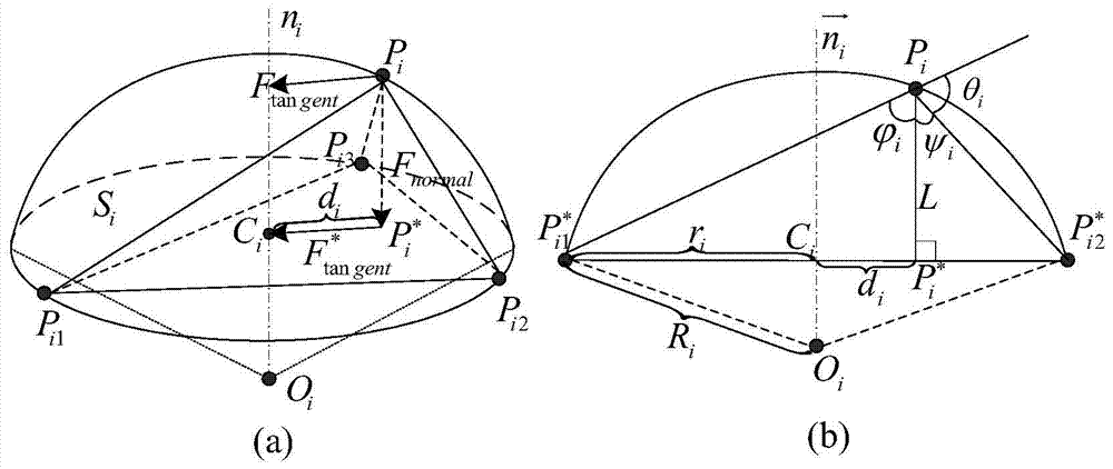
[0030] as attached figure 2 As shown, the internal force of the model is divided into tangential force F tangent a...


PUM


Abstract

The invention provides a self-adaptive surface deformation model based CT (Computed Tomography) image liver segmentation method and a convenient tool is provided for the clinical diagnosis of liver diseases. The self-adaptive surface deformation model based CT image liver segmentation method comprises step 1, performing preprocessing on an image by an anisotropic filtering method to obtain an initial boundary image; step 2, describing the initial contour of the liver through a DSM (Deformable Simplex Model); step 3, calculating the internal force of the model according to the relation between a vertex of the model and the neighborhood of the vertex and calculating the external force of the model through the gradient and the boundary of the original image; step 4, building a constraint model of the internal force and the external force of the model; step 5, performing self-adaptive decomposition of a triangular mesh; step 6, setting the number of iterations and approximating a target area under the driving of the internal force, the external force and the balloon force; step 7, obtaining an accurate segmentation result.

Description

technical field [0001] The invention relates to a CT image liver segmentation method based on an adaptive surface deformation model, belonging to the image scanning field of liver segmentation. Background technique [0002] The liver is the largest metabolic organ in the human body, the synthesis, decomposition, transformation and storage center of the substances and nutrients needed by the human body. Liver segmentation is an important basis for computer-aided diagnosis and treatment, tumor resection, surgical planning and living donor transplantation. The result of liver segmentation directly affects the accuracy of liver three-dimensional reconstruction, and affects doctors' diagnosis and treatment of liver lesions. Therefore, the research on liver segmentation method not only has important academic significance but also has important clinical application value. [0003] However, in computed tomography (CT) images, the gray value of the liver tissue is often similar to ...

Claims


Application Information

Patent Timeline
Patent Type & Authority Patents(China)
IPC IPC(8): G06T7/149
CPCG06T7/12G06T2207/10081G06T2207/20112G06T2207/30056
Inventor 杨健王雪虎王涌天刘越艾丹妮
Owner ARIEMEDI MEDICAL SCI BEIJING CO LTD
Learn More