
Method for internal compression hemostasis after minimally invasive breast lump rotary varicotomy and hemostatic balloon

A breast lump, compression hemostasis technology, applied in the direction of surgery, medical science, etc., can solve the problems of gauze and chest belt displacement, rebleeding in tissue defect areas, hemorrhagic shock of patients, etc., to achieve accurate position, durable and reliable hemostasis effect Effect

Inactive Publication Date: 2015-06-10
THE OBSTETRICS & GYNECOLOGY HOSPITAL OF FUDAN UNIV
View PDF10 Cites 1 Cited by
  • Summary
  • Abstract
  • Description
  • Claims
  • Application Information

AI Technical Summary

Problems solved by technology

However, the gauze and the chest strap will also shift when the patient wears a chest strap during minor daily activities. At this time, rebleeding will occur in the tissue defect area, and then a huge hematoma will form, and the bleeding volume will sometimes reach hundreds of milliliters. even lead to hemorrhagic shock

Method used


Image

  • Method for internal compression hemostasis after minimally invasive breast lump rotary varicotomy and hemostatic balloon

Examples


Embodiment Construction

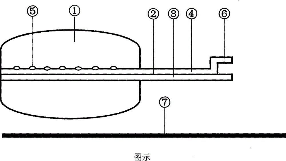
[0009] The present invention consists of three parts: a liquid-fillable balloon ①, a double-lumen catheter ② and a catheter core ⑦. The front end of the injection catheter lumen ④ is located in the balloon, and there is a side hole ⑤ in it, and the end is equipped with a self-closing liquid injection cap ⑥; the front end of the drainage catheter lumen ③ is flush with the front end of the balloon, and the end is open . The above-mentioned main structure is made of medical silica gel or medical rubber. Catheter core ⑦ is made of polyethylene material, should have certain hardness and elasticity, and its diameter is slightly smaller than the diameter of drainage catheter lumen ③, and length is about 3 centimeters longer than the latter.

[0010] After minimally invasive rotary breast mass resection, insert the catheter core ⑦ into the drainage catheter lumen ③. At this time, the fluid-inflatable balloon ① is still in a non-filled state, and the front end of the double-lumen cath...


PUM


Abstract

The invention discloses a method for internal compression hemostasis after minimally invasive breast lump rotary varicotomy and a hemostatic balloon. A tissue defect area caused by the minimally invasive rotary varicotomy can be effectively and reliably filled, and is internally compressed for hemostasis instead of the conventional common external compression hemostasis method, and reliable and long-lasting hemostasis effects are achieved. The hemostatic balloon comprises three parts, namely a liquid-filled balloon 1, a double-cavity catheter 2 and a catheter core 7. After the minimally invasive breast lump rotary varicotomy, the catheter core 7 is arranged in a drainage catheter cavity 3, the front end of the double-cavity catheter 2 is arranged in an operative incision, and reaches the tissue defect area, and when the balloon 1 is observed and regulated into a satisfied depth by virtue of B ultrasonic examination, the balloon 1 is filled with normal saline through a liquid filling catheter cavity 4 until the tissue defect area is completely compressed. After an injector is removed, a self-closing liquid filling cover 6 is closed, and the balloon 1 is in a continuously filled state. A small amount of normal saline seeps, and can flow out of the body through the drainage catheter cavity 3. When the injector is removed, the normal saline in the balloon 1 is sucked out until the balloon 1 completely returns to a preoperative state.

Description

technical field [0001] The invention relates to an internal compression hemostasis method after minimally invasive rotary excision of a breast mass and a hemostatic balloon used for compression hemostasis. Using the hemostatic balloon for internal compression hemostasis can effectively and accurately fill the tissue defect area caused by the operation, and the hemostatic effect is reliable. And lasting. technical background [0002] After minimally invasive rotary breast lumpectomy, a closed tissue defect will be formed. There will be hemorrhage in this area, and many patients will experience active bleeding. Because the bleeding cannot be stopped under direct vision, continuous pressure bandaging is required to achieve hemostasis the goal of. In the previous clinical work, the method used was: cover the skin surface of the tissue defect with multiple layers of gauze, and then bandage the chest belt with continuous pressure. This is a typical external compression hemostasis...

Claims


Application Information

Patent Timeline
Patent Type & Authority Applications(China)
IPC IPC(8): A61B17/12
CPCA61B17/12013A61B2017/12004
Inventor 宋晖
Owner THE OBSTETRICS & GYNECOLOGY HOSPITAL OF FUDAN UNIV
Learn More