A method of detecting quantum dot-protein binding kinetics

A quantum dot and kinetic technology, applied in the direction of electrochemical variables of materials, etc., can solve the problems of difficult separation of quantum dot-protein complexes, low resolution, etc.

Inactive Publication Date: 2015-07-08
CHANGZHOU UNIV
View PDF1 Cites 5 Cited by
  • Summary
  • Abstract
  • Description
  • Claims
  • Application Information

AI Technical Summary

Problems solved by technology

At present, there are few studies on the binding kinetics of quantum dots-proteins, including fluorescence spectroscopy methods, gel chrom

Method used


Image

  • A method of detecting quantum dot-protein binding kinetics
  • A method of detecting quantum dot-protein binding kinetics

Examples


Embodiment 1

[0020] Detection of dBSA and QDs Kinetics in Capillary

[0021] 1. Fat-soluble QDs are converted into water-soluble QDs by GSH

[0022] Mix 18 mg GSH, 5 mg KOH, and 250 μL methanol, take 40 μL of the mixed solution and add it to 200 μL fat-soluble quantum dots, and shake for 30 minutes. After shaking, 200 μL of 1 mM NaOH was added, and the fat-soluble quantum dots were transferred to the water phase. The quantum dots in the upper layer were taken out, precipitated by adding 1 mL of methanol and 30 μL of NaCl solution (30 mg / mL), and dissolved in a pH 7.4 boric acid buffer. The precipitation was repeated twice, and finally dissolved in 200 μL pH7.4 boric acid buffer. The concentration of QDs does not change after switching.

[0023] 2. Preparation of dBSA solution

[0024] dBSA solution through NaBH 4 Prepared by reacting with BSA. The main steps are as follows: 0.33g BSA was dissolved in 100mL pH 7.4 borate buffer, then 0.008g NaBH was added while stirring 4 , stirred a...

Embodiment 2

[0030] Extracapillary detection of dBSA and QDs kinetics

[0031] Steps 1 and 2 are the same as in Example 1.

[0032] 3. QDs coupled with dBSA

[0033] Different ratios of dBSA and QDs were self-assembled outside the capillary for 60 s, and then injected for 20 s and detected by electrophoresis.

[0034] 4. Fluorescence capillary electrophoresis analysis and detection

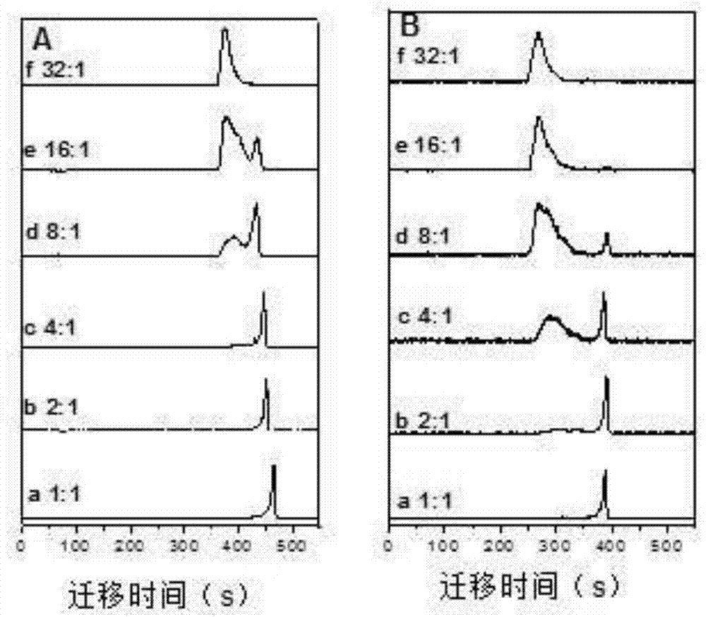
[0035] When we detected outside the capillary, with the increase of dBSA concentration, the electrophoretic peak of QDs gradually decreased, and the electrophoretic peak of the complex gradually increased, and the reaction was complete at 16:1 ( figure 1 B). According to S complex / S total and dBSA concentration, QDs and dBSA kinetic curves were obtained by Hill equation fitting ( figure 2 , curve b). K is calculated by the Hill equation D 2.8×10 -6 M, n is 2.8, R 2 is 0.994.


PUM


Abstract

The invention relates to the technical field of nanometer biology and particularly relates to a method of detecting quantum dot-protein binding kinetics. According to a technical scheme, the method researches the binding kinetics between quantum dots and protein through fluorescent capillary electrophoresis and fits a binding kinetic curve by the hill equation. By adoption of the technical scheme, the beneficial effects of the method are that the method is suitable for research on binding kinetic changes of the quantum dots and the protein outside and inside capillaries, broadens the application range of capillary electrophoresis, and provides theoretical bases for further application of quantum dot biological probes.

Description

technical field [0001] The invention relates to the field of nanobiology technology, in particular to a method for detecting quantum dot-protein binding kinetics. Background technique [0002] Quantum dots (QDs), as a novel fluorescent material, have the advantages of broad excitation spectrum, narrow and symmetrical emission spectrum, tunable color, anti-photobleaching and long fluorescence lifetime. These superior properties make QDs widely used in cell imaging and biomarking, and gradually become a very important probe tool in cell imaging research. [0003] At present, fat-soluble CdSe / ZnS quantum dots synthesized by metal-organic solvent method are the most widely used in biomarkers. Therefore, lipid-soluble quantum dots can be converted into water-soluble quantum dots by hydrophilic ligand exchange, amphiphilic ligand encapsulation, silane coating, and other methods. Water-soluble quantum dots can be used to prepare quantum dot fluorescent probes with proteins throug...

Claims


Application Information

Patent Timeline
IPC IPC(8): G01N27/26
Inventor 王建浩李静燕蒋鹏举邱琳李进晨王车礼秦玉琴柳丽滕一万
Owner CHANGZHOU UNIV
Why Eureka
Try Eureka