Medical image segmentation method and medical image segmentation device

A medical image and summation technology, applied in the field of medical image processing, can solve problems such as low efficiency and inconvenient use for users, and achieve the effect of high accuracy, robustness, and strong information integrity

Active Publication Date: 2015-11-25
SHANGHAI UNITED IMAGING HEALTHCARE
View PDF3 Cites 5 Cited by
  • Summary
  • Abstract
  • Description
  • Claims
  • Application Information

AI Technical Summary

Problems solved by technology

However, this method needs to pre-define the seed points of different bones. For the segmentation of a single bone, it is necessary to traverse the seed points of the surrounding bones, which is inefficient and inconvenient for users.

Method used


Image

  • Medical image segmentation method and medical image segmentation device
  • Medical image segmentation method and medical image segmentation device
  • Medical image segmentation method and medical image segmentation device

Examples


Embodiment Construction

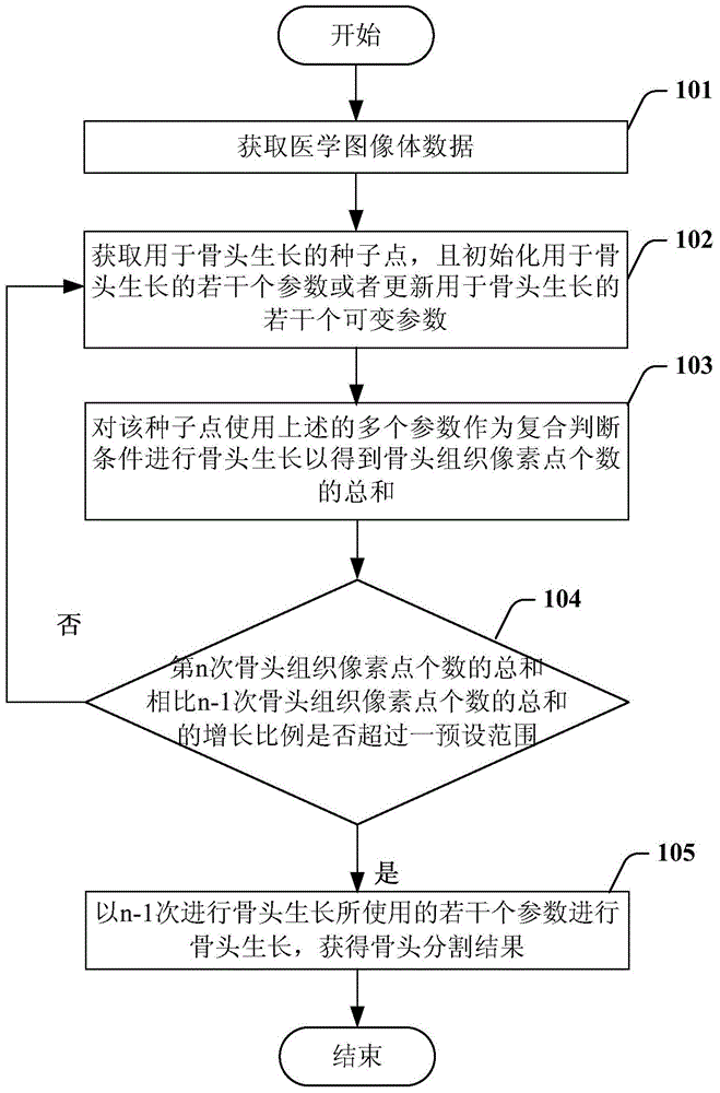
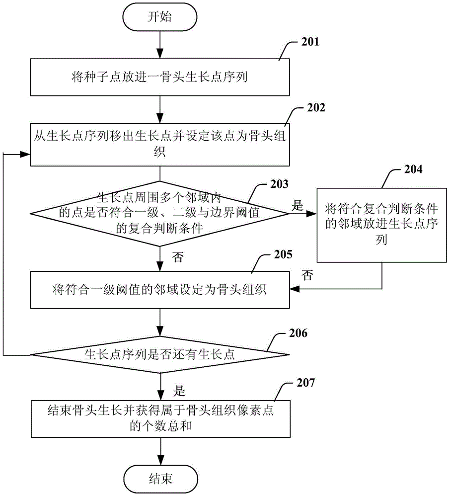
[0023] Embodiments of the present invention describe a medical image segmentation method and apparatus for segmenting bone regions from CT images. In the context of the present invention, the word "bone" refers to a bony region in an image, if not specified otherwise. In addition, it should be noted that the present invention can also be applied to MR images, XR images and other medical images.

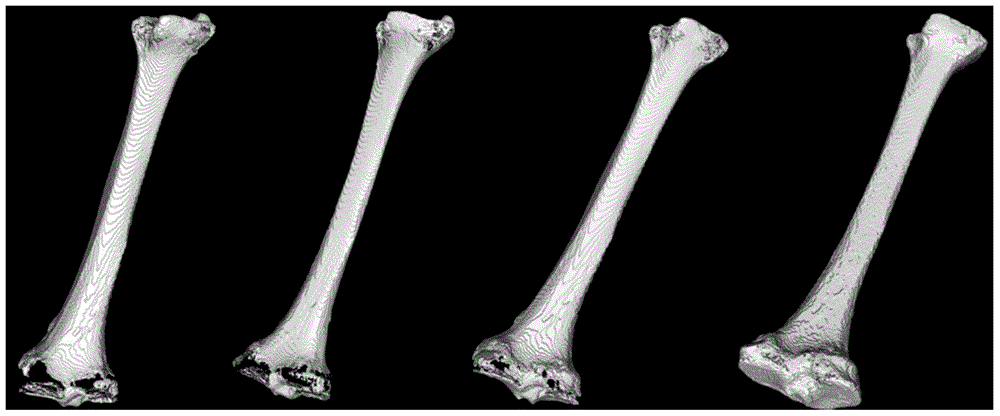
[0024] In a general CT image, the gray value of the bone is in a higher range and the range is larger. Accurately extracting bones has many benefits for the reconstruction of 3D information and medical diagnosis, such as viewing of bone articular surfaces, detection of the state of bone joint connections, and removal of bone information in CT images.

[0025] In summary, the embodiment of the present invention is a bone cutting method based on a single growth point, which separates the bone to which the seed point belongs, and is also effective for images with blurred boundaries and ...


PUM


Abstract

The invention relates to a medical image segmentation method and a medical image segmentation device which are used to segment a bone from a medical image. The method is characterized by comprising the following steps: parameters for bone growth are set, including a first-level threshold, a second-level threshold and a boundary threshold, wherein the first-level threshold is a fixed parameter, and the second-level threshold and the boundary threshold are variable parameters; bone growth is carried out on a seed point by taking the first-level threshold, the second-level threshold and the boundary threshold as composite judging conditions to obtain the total number of bone tissue pixels; and whether the bone tissue growth rate is beyond a preset range is judged, bone growth is iterated multiple times until optimized parameters are obtained if the bone tissue growth rate is not beyond a preset range, and final bone growth is carried out to obtain a bone segmentation result.

Description

technical field [0001] The present invention mainly relates to medical image processing, in particular to a medical image segmentation method and device. Background technique [0002] Computerized Tomography (abbreviated as CT) is a radiological diagnostic technique based on the fact that different substances have different attenuation properties for rays. CT irradiates the measured object with radiation from various directions, measures the intensity of the radiation passing through the object, and calculates the linear attenuation coefficient of each point inside the object for the radiation through a certain reconstruction algorithm, so as to obtain the radiological diagnosis of the tomographic image of the measured object technology. The tomographic image reconstructed by CT has the advantages of no image overlap, high density and high spatial resolution, so it has attracted much attention as a medical non-invasive diagnostic technology since its appearance. [0003] C...

Claims


Application Information

Patent Timeline
IPC IPC(8): G06T7/00
CPCG06T7/0012G06T2207/10081G06T2207/30008
Inventor 马延杰李程
Owner SHANGHAI UNITED IMAGING HEALTHCARE
Why Eureka
Try Eureka