Method, device and equipment for automatically removing ultrasound contrast imaging non-contrast region

A technology of contrast-enhanced ultrasound and imaging area, which is applied in the directions of ultrasound/sound wave/infrasonic wave diagnosis, sound wave diagnosis, infrasonic wave diagnosis, etc. It can solve time-consuming problems and achieve an effect that is conducive to observation

Inactive Publication Date: 2015-12-02
SONOSCAPE MEDICAL CORP
View PDF4 Cites 3 Cited by
  • Summary
  • Abstract
  • Description
  • Claims
  • Application Information

AI Technical Summary

Problems solved by technology

This requires a frame-by-frame operation of

Method used


Image

  • Method, device and equipment for automatically removing ultrasound contrast imaging non-contrast region
  • Method, device and equipment for automatically removing ultrasound contrast imaging non-contrast region
  • Method, device and equipment for automatically removing ultrasound contrast imaging non-contrast region

Examples


Embodiment 1

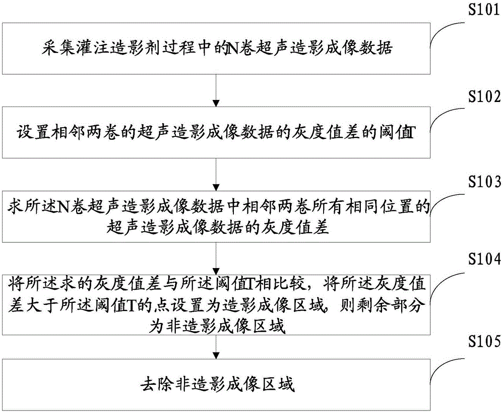
[0032] Such as figure 1 As shown, it is a method for automatically removing non-contrast areas of contrast-enhanced ultrasound imaging according to a specific embodiment of the present invention, which specifically includes the following steps:

[0033] S101: Collect N-volume ultrasound contrast imaging data during the process of perfusing the contrast agent.

[0034] The ultrasound contrast imaging data of the same part of the contrast agent perfusion process is collected through the probe.

[0035] The probe ultrasound transducer transmits ultrasound signals to the target tissue and receives echo signals. The echo signal is converted into an analog electric signal, and finally the analog electric signal is converted into a digital signal through an A / D conversion chip. The digital signal here is the ultrasound contrast imaging data.

[0036] Each time the probe is scanned, a volume of contrast-enhanced ultrasound data is obtained. During the process of contrast agent perfusion, the...

Embodiment 2

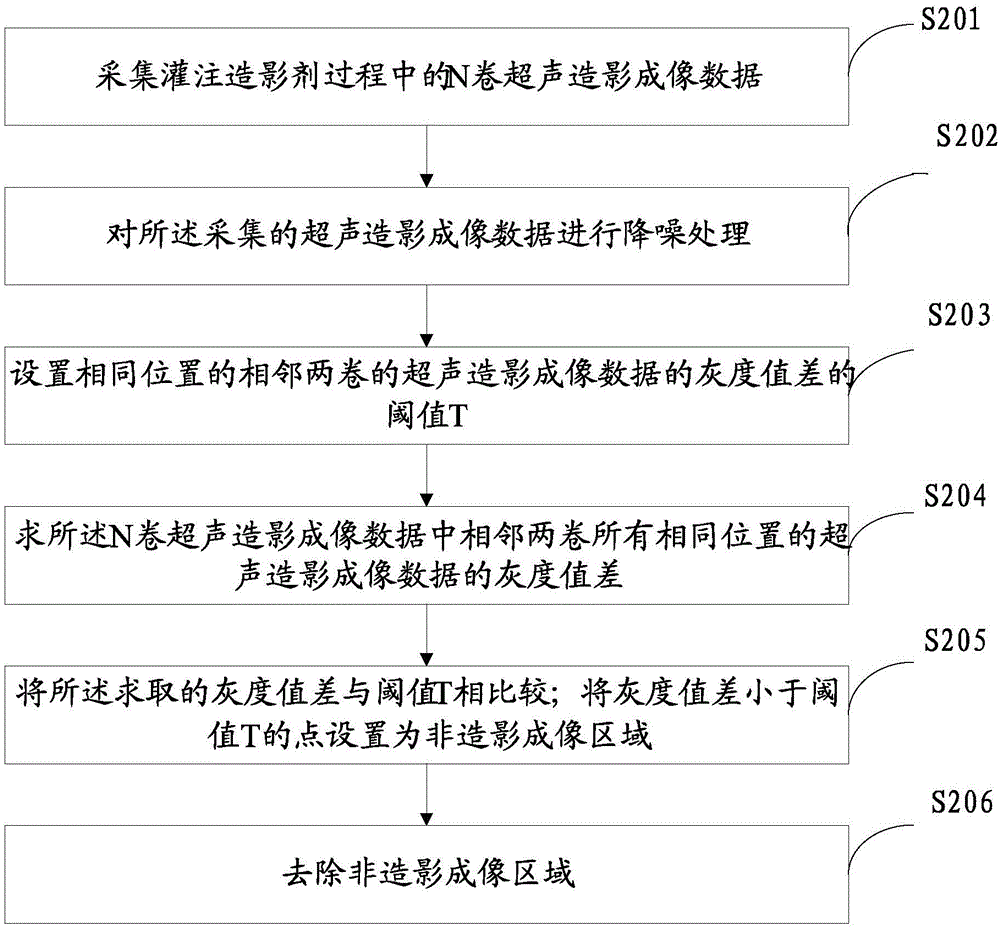
[0071] Such as Figure 4 As shown, the specific embodiment of the present invention also provides an apparatus 300 for automatically removing the non-contrast area of ​​ultrasound contrast imaging. The device includes:

[0072] The acquisition unit 301 is used to acquire N-volume ultrasound contrast imaging data during the process of contrast agent perfusion.

[0073] The first calculation unit 302 is used for the threshold value T of the gray value difference of the contrast-enhanced ultrasound imaging data of two adjacent volumes;

[0074] The second calculation unit 303 is configured to calculate the gray value difference of the ultrasound contrast imaging data at all the same positions in two adjacent volumes of the N volumes of ultrasound contrast imaging data;

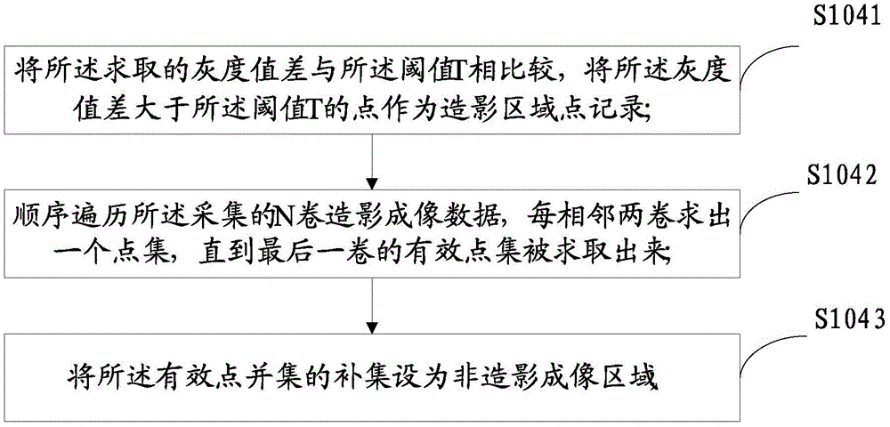
[0075] The third calculation unit 304 is configured to compare the obtained gray value difference with the threshold value T, and set the point where the gray value difference is greater than the threshold value T as the...

Embodiment 3

[0084] It is a device according to an embodiment of the present invention. The device is an ultrasonic device, and the ultrasonic device includes the device described in any of the second embodiment. For the device, refer to the second embodiment, and will not be repeated here.


PUM


Abstract

The invention provides a method for automatically removing an ultrasound contrast imaging non-contrast region. The method comprises the following steps: collecting N rolls of ultrasound contrast imaging data in a filling process of a contrast agent, wherein N is greater than or equal to 2; setting a grey value difference threshold T of two adjacent rolls of ultrasound contrast imaging data; solving a grey value difference between every two adjacent rolls of ultrasound contrast imaging data at all the same positions in the N rolls of ultrasound contrast imaging data; comparing the solved grey value difference with the threshold T, setting a point where the grey value difference is greater than the threshold T as a contrast imaging region, estimating a background region by solving a complementary set of N rolls of contrast imaging region union sets, and removing the background region. The invention also provides a corresponding device and corresponding equipment. By the adoption of the method, the device and the equipment, the background region is estimated according to the grey value difference between every two adjacent rolls of data at the same positions in the filling process of the contrast agent and is removed for each roll of data of an ultrasound image, so that the operation trouble of manually removing the background region by a doctor in ultrasound contrast imaging is eliminated.

Description

Technical field [0001] The present invention relates to the technical field of ultrasound imaging, in particular to a method, device and equipment for automatically removing non-contrast areas of ultrasound contrast imaging. Background technique [0002] Contrast-enhanced hysterosalpingist four-dimensional imaging uses a motor-driven volume probe to collect multi-volume contrast volume data, and present the flow and diffusion of the contrast agent in the uterus, fallopian tubes, and pelvis in a three-dimensional three-dimensional movie (four-dimensional) come out. In this process, the doctor diagnoses the patient's physiological condition based on the flow pattern of the contrast agent, the overflow pattern of the contrast agent at the fimbriae of the fallopian tube, and the diffuse pattern of the contrast agent in the pelvic cavity when it exits the fimbria of the fallopian tube. [0003] Due to the influence of ultrasound image noise, artifacts, and tissue echo signals, the imag...

Claims


Application Information

Patent Timeline
IPC IPC(8): A61B8/00
Inventor 黄柳倩孙慧刘旭江
Owner SONOSCAPE MEDICAL CORP
Why Eureka
Try Eureka