Clustered volley method and apparatus

A clustering, pupillary technique applied in the field of assessing the operation of the visual sensory system, capable of solving problems such as non-existence

Active Publication Date: 2015-12-30
KONAN MEDICAL USA
View PDF9 Cites 5 Cited by
  • Summary
  • Abstract
  • Description
  • Claims
  • Application Information

AI Technical Summary

Problems solved by technology

Responses to stimuli can be detected by monitoring the pupil, which has the advantage of allowing non-contact assessment, but until now there has been no commercial perimetry system using a pupillometer for multifocal testing of the nervous system

Method used


Image

  • Clustered volley method and apparatus
  • Clustered volley method and apparatus
  • Clustered volley method and apparatus

Examples


example 1

[0100] Example 1 - Improved SNR for normal subjects

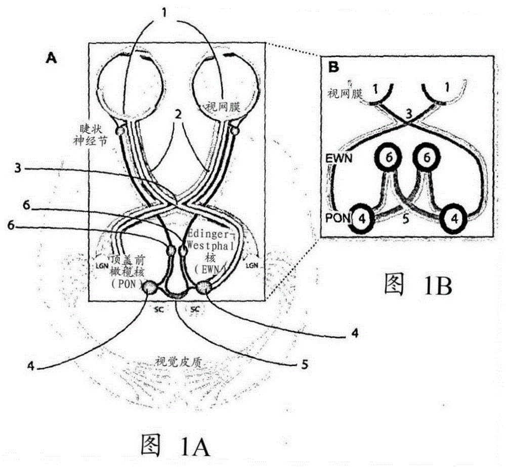
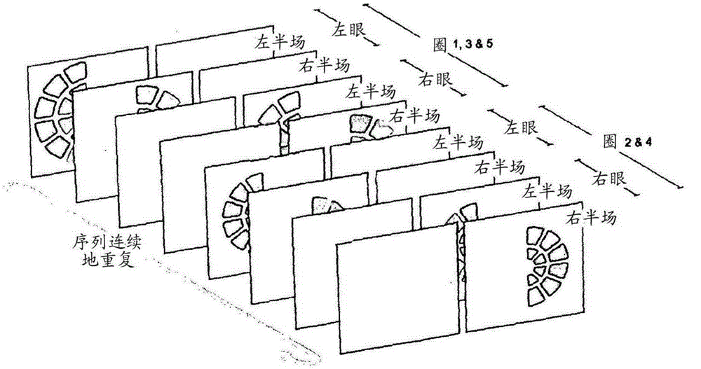
[0101] International PCT Patent Publication WO / 2005 / 051193 describes a spatially sparse multifocal stimulation type. The idea disclosed in this document is that there is a gain control operation within the retinal part of the visual nervous system. Most of the sensory parts of the brain lie on sheets of neural tissue that are often folded to fit within the skull, forming similar gyri and sulci to the human brain. The sensory part of the brain with retinal re-presentation contains a sensory brain map of the visual field on its surface. Other types of sensory fields were also mapped in different parts of the brain slice. Within this re-rendering, there is the possibility that the gain control can interact on-chip in a short range. Retinal mapping implies that average gain control can affect responses to stimuli in adjacent regions of the visual field. International PCT Patent Publication WO / 2005 / 051193 shows that such reg...

example 2

[0122] Example 2 – Clustered salvo stimuli in glaucoma

[0123] Investigate the relative diagnostic efficacy for distinguishing normal subjects from patients with open-angle glaucoma. The three wide-area stimuli in Table 3, namely Yold, Ynew and RGnew, are 150cd / m 2 variable. As before, all three stimulation types had an average stimulation interval of 4 seconds per field. Use FA-II achromatic visual field examination (SITA-FAST), matrix 24-2 visual field examination, Heidelberg spectral domain optical coherence tomography (OCT), slit lamp biomicroscopy, and applanation tonometry to confirm the subject's diagnostic status. Subjects were required to pass the manufacturer's criteria for false positives, negatives, and loss of fixation on visual field examination. Exclusion criteria for all subjects included acuity below 6 / 12, distance refraction greater than ±6 diopters or greater than 2 diopters of cylinder. Normal subjects have little to no relation to glaucoma. The study...

example 3

[0136] Example 3 – Clustered salvo stimulation in macular degeneration

[0137] Versions of the three macular-directed stimulus types using Table 4 to compare the diagnostic efficacy of old spatially sparse stimuli and SteadyOld for the detection of age-related macular degeneration (AMD), with two new clustered stimuli FlickNew and SteadyNew, of which 44 Stimulus areas were presented to the central 30 degrees of each field of view. Stimuli are yellow, similar to Form 4, but have a maximum brightness of 288cd / m in their active state 2 , present at 10cd / m 2 on the background. Table 8 summarizes the patient and age-matched normal control subject statistics. All subjects were examined for the glaucoma study above, exclusion criteria were similar. In addition, color retinal pictures were taken using a non-mydriatic fundus camera with a 45-degree field of view.

[0138] Form 8

[0139] subject group

number of subjects

number of men

Age±SD years

norm...


PUM


Abstract

Systems and methods are disclosed for assessing the function of parts of one or more sensory fields of a subject. Pupillary responses to at least two clustered ensembles of stimuli presented to predetermined portions of the sensory fields to be tested are measured. Each cluster comprises individual stimuli presented at locations across the sensory field, where the locations are defined on appropriate axes for the tested sensory fields. The method comprises: presenting statistically independent sequences of selected individual stimuli from the two or more clustered stimulus ensembles to a sensory field of a subject, thereby evoking pupillary responses in at least one pupil of the subject; detecting responses of the pupil or pupils evoked by the stimuli using at least one sensor; and processing the detected responses to relate the detected response to the sensory function of each component part of the sensory field. The sensory fields may be, but are not limited to, the visual fields of the two eyes of a subject.

Description

[0001] related application [0002] This application claims the benefit of an earlier filing date of Australian Provisional Patent Application No. 2012905171, filed 26 November 2012, with the same title as this application, which application is hereby incorporated by reference in its entirety. technical field [0003] The present invention relates generally to methods and systems for assessing the function of the nervous system using the pupil and unique properties of the pupil, and in particular to methods and systems for assessing the operation of the visual sensory system. Background technique [0004] Any discussion of background art throughout this specification should not be construed as an admission that such background art is widely known or forms part of the common general knowledge in the field. [0005] In humans, each optic nerve contains approximately 1,000,000 nerve fibers that carry information from all parts of each retina to the brain. An important design f...

Claims


Application Information

Patent Timeline
Patent Type & Authority Applications(China)
IPC IPC(8): A61B3/10
CPCA61B3/024A61B3/032A61B5/1104A61B5/4005A61B3/0025A61B3/0041A61B3/112A61B3/113A61B3/1173
Inventor 特迪·莉·麦蒂斯安德鲁·查尔斯·詹姆士科琳·弗朗西斯·卡尔斯
Owner KONAN MEDICAL USA
Why Eureka
Try Eureka