

Metal ion reagents and imaging preparations for early and rapid detection of malignant tumors and cardiovascular and cerebrovascular related diseases and multimodal imaging
A multi-modal imaging and metal ion technology, applied in the field of medical imaging, can solve the problems of poor targeting of preparations, weak signal strength, and high toxicity in vivo, and achieve simple and easy detection methods, avoid biological toxicity, and high sensitivity Effects of rapid tracing and monitoring
- Summary
- Abstract
- Description
- Claims
- Application Information
AI Technical Summary
Problems solved by technology
Method used
Image


Examples
Embodiment 1
[0029] 1. Preparation of metal ion reagents
[0030] Preparation of metal ion reagents with excellent biocompatibility, the specific steps include:
[0031] The compound zinc chloride and europium sulfate were dissolved in ultrapure water respectively, and were prepared into a solution with a concentration of 10mmol / L; after mixing the zinc chloride solution and europium sulfate in equal proportions, they were mixed with 10mmol / L ferric chloride aqueous solution (chlorine Dissolve ferric oxide in ultrapure water) and mix according to volume 1:1 to obtain metal ion reagent.
[0032] 2. In vitro test
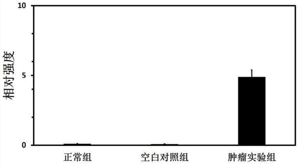
[0033]Application of the metal ion reagent of this embodiment to body fluid (such as blood or serum, etc.) or urine was tested, and the results showed that the optical and electrochemical properties of the tumor experimental group changed significantly after the metal ion reagent was added, while the normal control group did not change significantly.
[0034] Among them, the bo...
Embodiment 2
[0054] 1. Preparation of metal ion reagents
[0055] Preparation of metal ion reagents with excellent biocompatibility, the specific steps include:
[0056] The compound zinc gluconate was dissolved in ultrapure water to prepare a solution with a concentration of 10mmol / L; the zinc gluconate solution was mixed with 10mmol / L ferrous chloride aqueous solution at a volume ratio of 1:3 to obtain a metal ion reagent.
[0057] 2. In vitro test
[0058] Metal ion reagents were used to test body fluids (such as blood or serum, etc.) or urine, and the results showed that the optical and electrochemical properties of the tumor experimental group changed significantly after the addition of metal ion reagents, while the normal control group had no significant changes .
[0059] Among them, the body fluid comes from normal people and leukemia patients, and the urine comes from normal nude mice and tumor model nude mice. Tumor models in nude mice were liver cancer (HepG2 cell line) and c...
Embodiment 3
[0070] 1. Preparation of metal ion reagents
[0071] Preparation of metal ion reagents with excellent biocompatibility, the specific steps include:
[0072] The compound chloroauric acid and silver nitrate were dissolved in ultrapure water respectively, and both were prepared into a solution with a concentration of 10mmol / L; after mixing the chloroauric acid solution and the silver nitrate solution in equal proportions, they were mixed with 10mmol / L ferric chloride aqueous solution according to The volume is mixed at 1:2 to obtain the metal ion reagent.
[0073] 2. In vitro test
[0074] Metal ion reagents were used to test body fluids (such as blood or serum, etc.) or urine, and the results showed that the optical and electrochemical properties of the tumor experimental group changed significantly after the addition of metal ion reagents, while the normal control group had no significant changes .
[0075] Among them, the body fluid comes from normal people and leukemia pa...
PUM
 Abstract
Description
Claims
Application Information