Medical ultrasound assisted automatic diagnosis device and medical ultrasound assisted automatic diagnosis method

An automatic diagnosis and ultrasound-assisted technology, which is applied in the directions of acoustic wave diagnosis, infrasonic wave diagnosis, ultrasonic/sonic wave/infrasonic wave diagnosis, etc., can solve problems such as heavy workload, uneven level of doctors, and influence on diagnosis, so as to improve efficiency and accuracy Effect

Inactive Publication Date: 2016-01-13
CHISON MEDICAL TECH CO LTD
View PDF9 Cites 24 Cited by
  • Summary
  • Abstract
  • Description
  • Claims
  • Application Information

AI Technical Summary

Problems solved by technology

[0004] The purpose of the present invention is to solve the problem that the inspection workload of glandular lesions in ultrasonic diagnosis is heavy, and the

Method used


Image

  • Medical ultrasound assisted automatic diagnosis device and medical ultrasound assisted automatic diagnosis method
  • Medical ultrasound assisted automatic diagnosis device and medical ultrasound assisted automatic diagnosis method
  • Medical ultrasound assisted automatic diagnosis device and medical ultrasound assisted automatic diagnosis method

Examples


Embodiment Construction

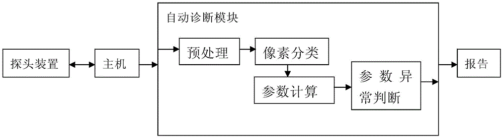
[0019] The device obtains the ultrasonic echo signal of the gland through the ultrasonic probe, and obtains the ultrasonic grayscale image for diagnosis through the signal detection, signal processing and image processing of the host computer. The grayscale image of the gland generated by the host computer is input to the automatic diagnosis module. In this module, the image is analyzed and processed by computer, and the result is transferred to the report module, and the detection report is output in accordance with the format required by the medical report.

[0020] The automatic diagnosis module includes several parts such as preprocessing, pixel classification, parameter calculation, and parameter abnormality judgment. The grayscale image transmitted from the host is subjected to noise reduction and enhancement processing in the preprocessing link, pixel classification is performed on the preprocessed image, the pixel data is divided into suspected lesion pixels and normal ...


PUM


Abstract

The invention relates to a medical ultrasound assisted automatic diagnosis device and a medical ultrasound assisted automatic diagnosis method. The method comprises the following steps of: obtaining an ultrasound echo signal of a human body tested position through an ultrasound probe; obtaining an ultrasound gray-scale image for diagnosis through host processing; inputting the ultrasound gray-scale image into an automatic diagnosis module; and performing computer analysis on the image by the automatic diagnosis module, wherein the computer analysis includes pixel classification, parameter calculation and parameter abnormality judgment; according to the pixel classification, pixel data of the image is divided into lesion suspected pixels and normal tissue pixels; according to the parameter calculation, lesion suspected pixels and peripheral pixels are subjected to geometrical and gray-scale relevant parameter calculation; and according to the parameter abnormality judgment, whether each geometrical and gray-scale relevant parameter is abnormal or not is judged, the disease degree is judged according to the abnormal condition of the geometrical or gray-scale relevant parameter, and a detection report is finally output. The device and the method have the advantages that an ultrasound image computer processing technology is used, and gland lesion assisted automatic diagnosis equipment is provided, so that the efficiency and the accuracy of the gland ultrasound examination performed by a user are improved.

Description

technical field [0001] The invention relates to a method and device for auxiliary diagnosis of ultrasonic equipment, which are used for auxiliary automatic diagnosis of pathological information such as glands, nerves, and blood flow. Background technique [0002] Breast, heart, liver and other diseases have become the diseases with higher cancer-causing rates in the world, and the incidence rate is increasing year by year and becoming younger. Compared with imaging diagnostic methods such as mammography, CT, and MRI, ultrasound imaging can clearly display glands, nerves, blood flow and other lesion information, such as breast and mass structures and lesions, and accurately identify cystic and solid lesions. With the advantages of dense breasts and no radiation, it is a relatively cheap, safe, and highly accurate examination method. Theoretically, the accuracy of ultrasonography can reach more than 96%, but general imaging examinations have higher requirements for doctors. In ...

Claims


Application Information

Patent Timeline
IPC IPC(8): A61B8/00
Inventor 王勇莫善珏赵明昌
Owner CHISON MEDICAL TECH CO LTD
Why Eureka
Try Eureka