Medical MR image segmentation method based on Hough transform and geometric active contour

An image segmentation and active contour technology, applied in the field of medical MR image segmentation, can solve problems such as smooth structure, high numerical calculation complexity, and not very ideal

Inactive Publication Date: 2016-01-13
NANNING BOCHUANG INFORMATION TECH DEV CO LTD
View PDF2 Cites 11 Cited by
  • Summary
  • Abstract
  • Description
  • Claims
  • Application Information

AI Technical Summary

Problems solved by technology

However, for images with uneven gray levels and noisy images, the performance of GVF will be affected, or only a suboptimal GVF field can be obtained, or the structure of the target may be smoothed
At this time, the evolution curve will enter another region from the weak boundary, causing boundary leakage or stopping evolution at the local maximum value of the gradient inside the region and the isolated edge, and cannot move to the real boundary.
Although the Paragios method adds the regional information obtained by fitting the histogra

Method used


Image

  • Medical MR image segmentation method based on Hough transform and geometric active contour

Examples


Embodiment Construction

[0017] This embodiment adopts the following technical solutions: it includes the following points:

[0018] 1. Using the prior shape knowledge that the inner and outer contours of the left ventricular myocardium in the short-axis image are approximately circular, the initial contour of the left ventricle is automatically positioned by Hough transform, so that the initial contour is more accurately positioned near the edge of the real contour, and then in the On the basis of the geometric active contour model, using the regional information provided by K-means clustering to roughly segment the target in the image and the physiological structure constraints of the myocardium, the coupled evolution equation of the inner and outer contour curves of the myocardium is established, and the left ventricular The inner and outer contours are automatically segmented at the same time.

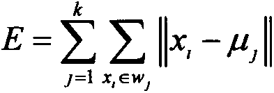
[0019] 2. K-means clustering algorithm

[0020] Clustering is to divide the data into multiple groups ...


PUM


Abstract

The present invention discloses a medical MR image segmentation method based on Hough transform and a geometric active contour and belongs to the technical field of image processing. According to the method, firstly, the prior shape knowledge that inner and outer contours of a left ventricle myocardium on a short axis image are of a round-like shaped is utilized, Hough transform is adopted to estimate an initial contour of a left ventricle and the reason of adopting Hough transform is that Hough transform has strong robustness, is not quite sensitive for the incompleteness of data or noise and can recognize partially deformed or partially shielded objects; the reason of using a K-means clustering algorithm is that as a square error based clustering method, the K-means clustering algorithm is simple and has a high clustering speed; and by using the method provided by the present invention, the inner and outer contours of the left ventricle can be segmented effectively, relative positions of evolving curves of the inner and outer contours of the left ventricle can be controlled and a function of shape constraint can be realized.

Description

Technical field: [0001] The invention relates to the technical field of image editing, in particular to a medical MR image segmentation method based on Hough transform and geometric active contour. Background technique: [0002] For the segmentation of left ventricular myocardium, the mainstream method is based on the edge-driven image segmentation method. Such as: parameter active contour model (also known as Snake model), level set (LevelSet) model, geometric active contour model and their improved models. However, mastoid muscle interference, local gradient maxima, weak edges, and artifacts often appear in MR images, which brings difficulties to edge-based image segmentation methods. Aiming at the problems caused by these phenomena, a lot of prior knowledge is introduced into the edge-based image segmentation model to improve the robustness of the model. The shape constraints and regional information based on prior knowledge are mostly used. Based on the geometric activ...

Claims


Application Information

Patent Timeline
IPC IPC(8): G06T7/00
CPCG06T7/0012G06T2207/10088G06T2207/20061G06T2207/30048
Inventor 纪东升王寿年廖开明
Owner NANNING BOCHUANG INFORMATION TECH DEV CO LTD
Why Eureka
Try Eureka