Dividing method based on CT image liver tumor focus

A technology for CT imaging and liver tumors, applied in the field of medical image processing, can solve the problems of inability to accurately and quickly realize the segmentation of multi-focal liver tumors, and achieve the effects of improving timeliness, adjustable weights, and improving accuracy

Active Publication Date: 2016-04-13
THE SECOND PEOPLES HOSPITAL OF SHENZHEN
View PDF2 Cites 14 Cited by
  • Summary
  • Abstract
  • Description
  • Claims
  • Application Information

AI Technical Summary

Problems solved by technology

[0005] The purpose of the present invention is to provide a method for segmenting liver tumor lesions based on CT images, aiming to solve the problem of inability to accurately and quickly realize the segmentation of multi-focal liver tumors

Method used


Image

  • Dividing method based on CT image liver tumor focus
  • Dividing method based on CT image liver tumor focus
  • Dividing method based on CT image liver tumor focus

Examples


Embodiment Construction

[0016] In order to make the technical problems, technical solutions and beneficial effects to be solved by the present invention clearer, the present invention will be further described in detail below in conjunction with the embodiments. It should be understood that the specific embodiments described here are only used to explain the present invention, not to limit the present invention.

[0017] An embodiment of the present invention provides a method for segmenting liver tumor lesions based on CT images, comprising the following steps:

[0018] S01. Preprocessing the initial CT image;

[0019] S02. According to the preprocessed CT image, the ROI selection of the suspected lesion is completed in an interactive manner;

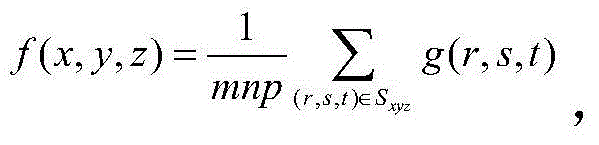
[0020] S03. Perform texture description on the ROI based on the CT value, and obtain the probability spectrum of the ROI through weighted calculation of the texture descriptor;

[0021] S04. Build a big data prior knowledge base, determine the threshold of ...


PUM


Abstract

The invention is applicable to the field of medical image processing, and provides a dividing method based on the CT (Computed Tomography) image liver tumor focus. The method comprises the following steps of preprocessing an initial CT image; completing ROI selection on the suspected focus in an interactive way by aiming at the preprocessed CT image; performing texture description on the ROI based on a CT value, and obtaining probability spectrums of the ROI through weighted computation of texture descriptors; building a big data priori knowledge base, determining a focus region probability spectrum threshold value, and dividing the suspected focus based on the threshold value; and completing the volume statistics and the quantization output on the focus. The method has the advantages that a plurality of texture descriptor probability spectrums are obtained based on stable and comparable CT value calculation; meanwhile, the threshold value of the texture descriptor probability spectrums is obtained through manual division calculation based on the priori knowledge base; and the accuracy and the timeliness of the multi-domain liver tumor focus division in the CT image can be realized.

Description

technical field [0001] The invention belongs to the field of medical image processing, in particular to a method for segmenting liver tumor lesions based on CT images. Background technique [0002] The liver is the largest chemical factory in the human body, undertaking important functions such as digestion, detoxification, and secretion. The burden of disease on the Chinese liver is the heaviest in the world. Primary liver cancer is one of the most common malignant tumors in clinic. At present, the incidence of liver cancer in the world is on the rise. According to the "Global Cancer Report 2014" published by the World Health Organization, China's new cancer cases rank first in the world, and the number of new cases and deaths of liver cancer ranks first in the world. At present, the incidence rate of liver cancer in my country is about 25.7 / 100,000, making it the third most deadly malignant tumor after gastric cancer and lung cancer. Early diagnosis of liver cancer is ...

Claims


Application Information

Patent Timeline
Patent Type & Authority Applications(China)
IPC IPC(8): G06T7/00
CPCG06T7/0012G06T2207/10081G06T2207/30056G06T2207/30096
Inventor 雷益王德峰黄俊彬李乐宁林帆
Owner THE SECOND PEOPLES HOSPITAL OF SHENZHEN
Why Eureka
Try Eureka