Brain ASL (Arterial Spin Labeling), SPECT (Single-Photon Emission Computed Tomography) and MRI (Magnetic Resonance Imaging) image registration and fusion conjoint analysis method and system

An image fusion and image technology, which is applied in the field of image processing and biomedical image processing, can solve the problems of image processing technology without fusion normalization analysis, image processing technology without MRI image joint analysis and processing, etc.

Inactive Publication Date: 2016-04-13
SHANGHAI JIAO TONG UNIV
View PDF5 Cites 26 Cited by
  • Summary
  • Abstract
  • Description
  • Claims
  • Application Information

AI Technical Summary

Problems solved by technology

At present, there is no image processing technology that completely integrates the two imaging methods and performs normalized analysis, and there is no

Method used


Image

  • Brain ASL (Arterial Spin Labeling), SPECT (Single-Photon Emission Computed Tomography) and MRI (Magnetic Resonance Imaging) image registration and fusion conjoint analysis method and system
  • Brain ASL (Arterial Spin Labeling), SPECT (Single-Photon Emission Computed Tomography) and MRI (Magnetic Resonance Imaging) image registration and fusion conjoint analysis method and system
  • Brain ASL (Arterial Spin Labeling), SPECT (Single-Photon Emission Computed Tomography) and MRI (Magnetic Resonance Imaging) image registration and fusion conjoint analysis method and system

Examples


Embodiment Construction

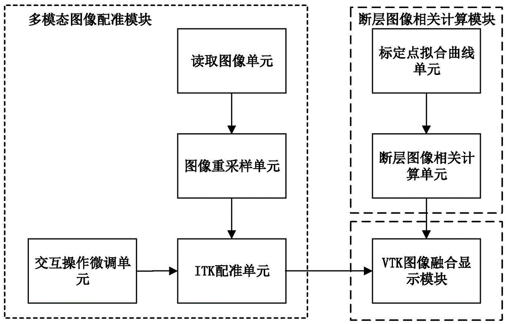
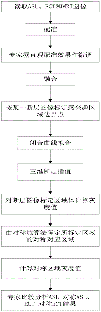
[0029] Such as figure 1 As shown, it is a system for marking pathological regions by fusion of brain ASL, SPECT and MRI images, including: VTK image fusion display module, multi-modal image auxiliary analysis composed of calibration point fitting curve unit and tomographic image correlation calculation unit module, and a three-modal image registration module composed of an image reading unit, an image resampling unit, an ITK registration unit, and an interactive fine-tuning unit, wherein: the three-modal image registration module is connected to the VTK image fusion display module, The data information of the three modal images after registration is displayed through the VTK image fusion display module; the multi-modal image auxiliary analysis module is connected with the VTK image fusion display module, and the area of ​​interest in the three modalities is displayed through the VTK image fusion display module The joint analysis calculation results.

[0030] The image reading...


PUM


Abstract

The invention discloses a brain ASL, SPECT and MRI image registration and fusion conjoint analysis method and system. Registration is respectively carried out to an ASL image and an MRI image by taking the MRI image as a standard; the ASL image and an SPECT image are fused according to different transparencies; a region to be computed is marked in the faultage of the SPECT image; a closed curve is obtained through carrying out a cubic spline interpolation to the ASL image; the volume and the gray value corresponding to the region to be computed and the gray value of an interesting region body through a circular truncated cone approach mode. According to the invention, the advantages of the ASL image reflected brain blood perfusion, the SPECT image reflected brain blood perfusion and the MRI provided brain structure change are combined; an interesting region is segmented and analyzed through providing interactive calibration operation a doctor and a medical expert; the judgement accuracy of a brain ischemic disease is improved; and iconography instructions are provided for a clinician to formulate a rational therapeutic schedule.

Description

technical field [0001] The present invention relates to a technology in the field of image processing, specifically a CBF (cerebral blood flow, Cerebral Blood Flow) measured by cerebral blood perfusion imaging of a brain SPECT (Single-PhotonEmission Computed Tomography, single photon emission computed tomography) A method for joint analysis of registration and fusion of ASL (Arterial Spin Labeling) images of imaging and MR (Magnetic Resonance, Magnetic Resonance) imaging belongs to the field of biomedical image processing, specifically a fusion display and display based on multimodal images. Techniques for Region of Interest Segmentation. Background technique [0002] Ischemic cerebrovascular disease is one of the diseases that seriously endanger human health. It has the characteristics of high morbidity, high mortality and high disability rate, and seriously affects people's quality of life. It is of great significance to detect the ischemic area early and provide the basi...

Claims


Application Information

Patent Timeline
IPC IPC(8): G06T7/00
CPCG06T2207/10088G06T2207/30101G06T2207/30104
Inventor 刘冶宋少莉王洋陈增爱王利生
Owner SHANGHAI JIAO TONG UNIV
Why Eureka
Try Eureka