
Method and system for a brain image pipeline and brain image region location and shape prediction

A brain image and assembly line technology, applied in the field of medical imaging systems, can solve problems such as lack of clear visualization, insufficient positioning, low therapeutic effect or side effects

Active Publication Date: 2016-06-01
オウルナビゲーションインコーポレイテッド
View PDF6 Cites 15 Cited by
  • Summary
  • Abstract
  • Description
  • Claims
  • Application Information

AI Technical Summary

Problems solved by technology

This is partly due to potentially lower therapeutic efficacy or side effects caused by insufficient positioning of the DBS electrodes in other areas of the STN (for PD) or other diseases
[0010] Manual segmentation is time consuming and largely driven by anatomical subjectivity due in part to the lack of clear visualization with clinical, often low-field MRI protocols

Method used


Image

  • Method and system for a brain image pipeline and brain image region location and shape prediction
  • Method and system for a brain image pipeline and brain image region location and shape prediction
  • Method and system for a brain image pipeline and brain image region location and shape prediction

Examples


Embodiment Construction

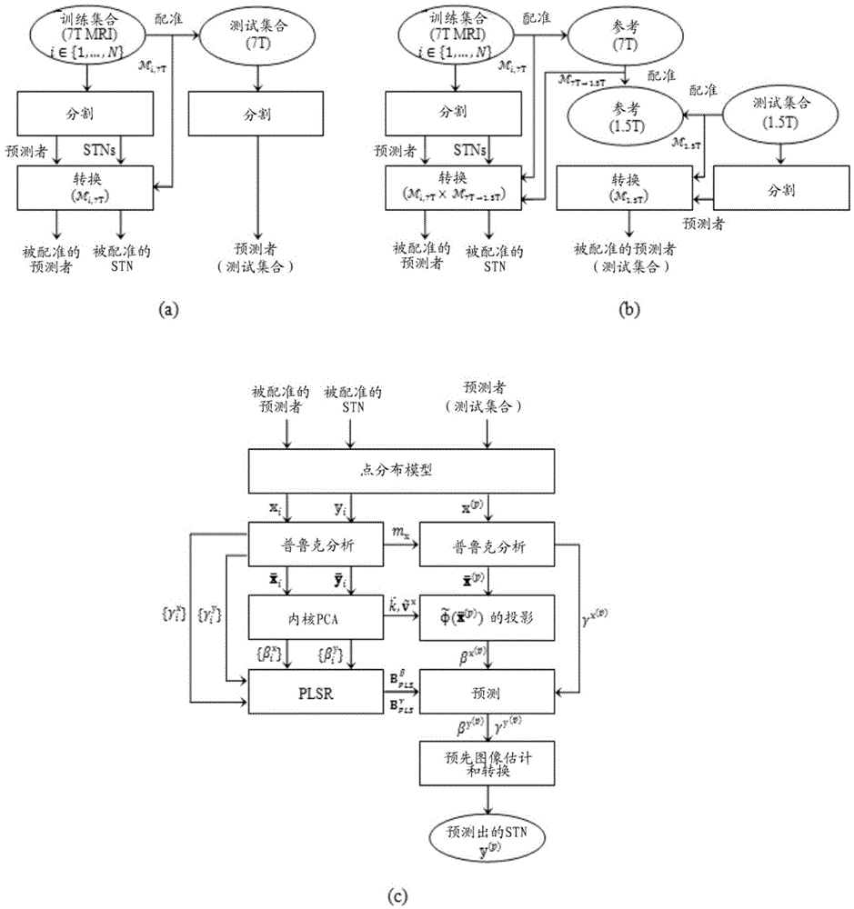
[0045]Recent advances in high-field MRI techniques enable direct in vivo visualization and localization of subcortical structures such as the STN and GPi, which is critical for DBS targeting for motion disorders such as PD. Visualization of these regions in three dimensions will provide surgical DBS targeting and postoperative programming more reliably and efficiently. However, it still has challenging issues: automatically delineate e.g. the STN region, since it is small and complex, there is unclear separation between adjacent structures such as SN. In particular, it is not possible to segment the STN in this straightforward manner when using images acquired according to standard low-field clinical protocols or when the data have a limited field of view within this region.

[0046] Methods for (high resolution) automatic shape prediction of STN or other brain structures relevant to brain operations are disclosed; STN is used here and throughout the document as a descriptive ...


PUM


Abstract

A volumetric segmentation method is disclosed for brain region analysis, in particular but not limited to, regions of the basal ganglia such as the subthalamic nucleus (STN). This serves for visualization and localization within the sub-cortical region of the basal ganglia, as an example of prediction of a region of interest for deep brain stimulation procedures. A statistical shape model is applied for variation modes of the STN, or the corresponding regions of interest, and its predictors on high-quality training sets obtained from high-field, e.g., 7T, MR imaging. The partial least squares regression (PLSR) method is applied to induce the spatial relationship between the region to be predicted, e.g., STN, and its predictors. The prediction accuracy for validating the invention is evaluated by measuring the shape similarity and the errors in position, size, and orientation between manually segmented STN and its predicted one.

Description

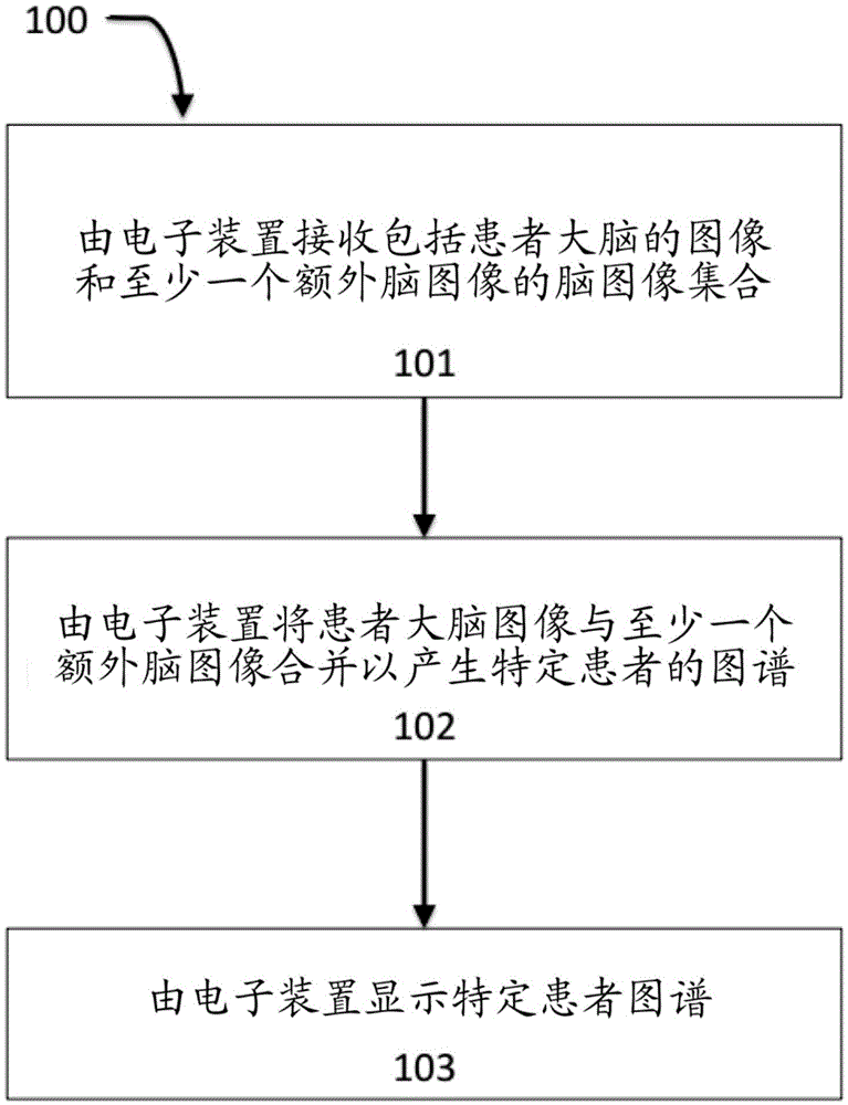
technical field [0001] Embodiments disclosed herein relate generally to medical imaging systems, and in particular to the generation and use of high-quality multimodal brain atlases of a specific patient in support of medical procedures, including but not limited to brain surgery and including deep brain stimulation (DBS). Background technique [0002] The use of medical images to guide neurosurgery is widespread. The brain is a complex organ, filled with structures and blood vessels that are both critical to a patient's survival and well-being, and difficult to perceive with imaging techniques alone. Three-dimensional imaging techniques that have become available, notably MRI using various contrast weights, have made individual internal features of the brain readily identifiable, ready for delicate, safe and more effective surgery. In practice, however, even a set of current imaging techniques, and especially those available in routine clinical settings, are insufficient t...

Claims


Application Information

Patent Timeline
IPC IPC(8): G06K9/54G06N20/00
CPCG06T2207/10088G06T2207/20128G06T2207/30016A61B6/032A61B6/037A61B6/12A61B6/469A61B6/501A61B6/5247A61B6/563A61B5/055A61B2576/026A61B5/0035G06T7/337G06T7/11G16H50/20G16H30/20G06N20/00G06F18/251G06F16/50A61B2034/107A61B6/5217A61B6/5294G06N5/04G06T7/0012G06T2207/10081
Inventor 吉列尔莫·萨皮罗诺姆·哈雷尔尤瓦尔·达钦金真英
Owner オウルナビゲーションインコーポレイテッド
Learn More