Multimode magnetic resonance imaging method for rat model under 3.0T field intensity

A magnetic resonance imaging and rat model technology, which is applied in the generation of 2D images, image enhancement, image analysis, etc., can solve the problems of inconvenience and lack of suitable structure for general products

Pending Publication Date: 2017-05-31
马国林 +1
View PDF1 Cites 4 Cited by
  • Summary
  • Abstract
  • Description
  • Claims
  • Application Information

AI Technical Summary

Problems solved by technology

[0004] It can be seen that there are obviously inconveniences and defects in the standardization and process of the above-mentioned existing brain functional magnetic resonance imaging scanning, diffusion tensor imaging scanning, and proton magnetic resonance spectroscopy scanning technology of the rat model under the 3.0T field strength. , and

Method used


Image

  • Multimode magnetic resonance imaging method for rat model under 3.0T field intensity
  • Multimode magnetic resonance imaging method for rat model under 3.0T field intensity
  • Multimode magnetic resonance imaging method for rat model under 3.0T field intensity

Examples


Embodiment Construction

[0027] For further elaborating the technical means and effects that the present invention takes to reach the intended purpose of the invention, below in conjunction with the accompanying drawings and preferred embodiments, the multimodal magnetic The specific implementation, features and effects of the resonance imaging method are described in detail below.

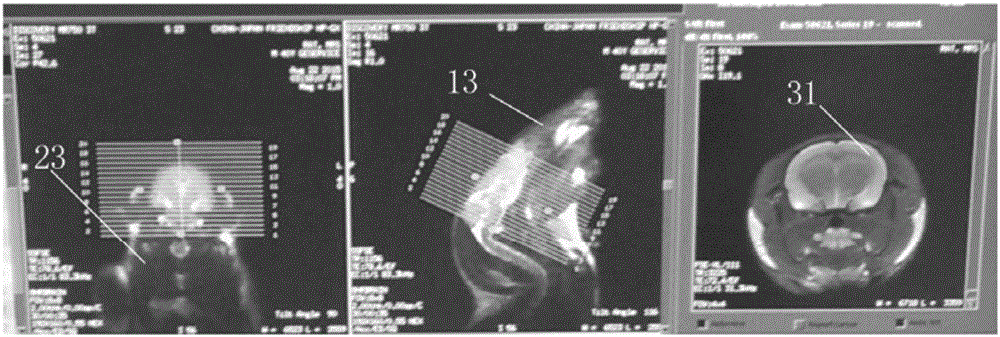
[0028] The manufacturer of the 3.0T field strength magnetic resonance scanner adopted in the present invention is General Medical (GE) Company of the United States, and the model is Discovery 750. The manufacturer of the 8-channel orthogonal rat coil is provided by Shanghai Chenguang Medical Technology Co., Ltd., and the model is CG-MUC30-H300-AG.

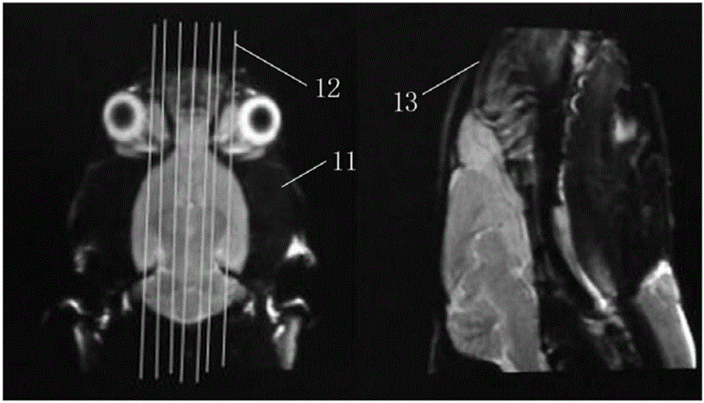
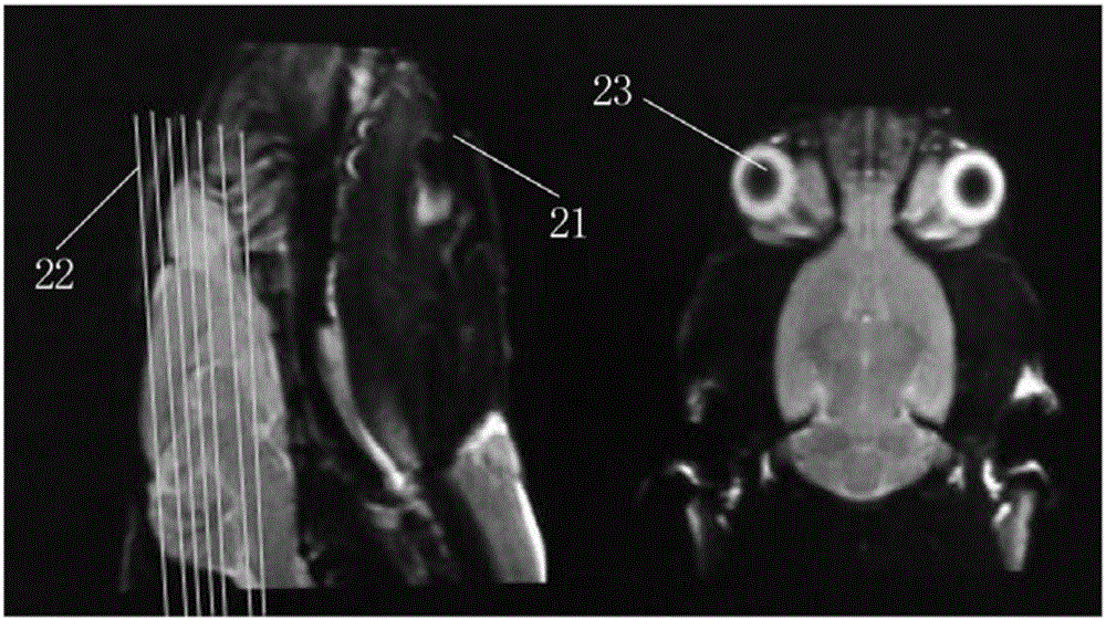
[0029] see Figure 1 to Figure 7 , the invention discloses a multi-modal magnetic resonance imaging method of a rat model under a 3.0T field strength, comprising the following steps:

[0030] S1, the rat model is anesthetized, and the rat model is fixed in a rat-specific coi...


PUM


Abstract

The invention discloses a multimode magnetic resonance imaging method for a rat model under 3.0T field intensity. The method comprises the following steps of S1, anesthetizing the rat model, and fixing the rat model in a special coil for a rat by adopting a prone position after anesthetization; S2, performing structure image scanning on the rat model; S3, performing brain function magnetic resonance imaging scanning on the rat model; S4, performing diffusion tensor imaging scanning on the rat model, wherein diffusion sensitive gradients are 32 different directions; and S5, finally performing proton magnetic resonance spectrum scanning on the rat model: determining a hippocampus and thalamus maximum layer in a coronal image subjected to visual field expansion, and taking the maximum layer as a scanning center layer, and then collecting a hippocampus and thalamus line two-dimensional multi-voxel spectrum of the rat model. By implementing the method, the brain function magnetic resonance imaging scanning, the diffusion tensor imaging scanning and the proton magnetic resonance spectrum scanning of the rat model under the 3.0T field intensity can be standardized, and a required clear scanning image can be obtained.

Description

technical field [0001] The invention relates to a tomography imaging method, in particular to a multimodal magnetic resonance imaging method of a rat model under a 3.0T field strength. Background technique [0002] At present, 3.0T magnetic resonance (MRI) scanners have been widely and commonly used in clinical and scientific research in hospitals. Due to their better soft tissue resolution, they can be well used in the research and diagnosis of central nervous system diseases. However, at present, the most applied models for studying animal models are ultra-high-field animal magnetic resonance scanners, ranging from 4.7T to 17.2T small animal MRI scanners. However, these ultra-high-field MRIs cannot enter hospitals for clinical diagnosis of patients in the short term. The reasons are as follows: On the one hand, the US FDA has not allowed any MRI above 7T to enter clinical practice. At the same time, the FDA approval department believes that MRI scans above 8.0T are harmfu...

Claims


Application Information

Patent Timeline
IPC IPC(8): G06T11/00G06T7/00A61B5/055
CPCG06T11/005A61B5/0042A61B5/055G06T7/0012G06T2207/10088
Inventor 马国林宋天彬
Owner 马国林
Why Eureka
Try Eureka