
Targeted nano-microbubbles for detection of small cell lung cancer, preparation method and application thereof

A small cell lung cancer, nanotechnology, applied in preparations for in vivo experiments, pharmaceutical formulations, echo/ultrasound imaging agents, etc.

Inactive Publication Date: 2018-10-19
SHANXI UNIV
View PDF5 Cites 0 Cited by
  • Summary
  • Abstract
  • Description
  • Claims
  • Application Information

AI Technical Summary

Problems solved by technology

[0005] At present, there is no research on the construction of nano-microbubbles targeted by anti-ProGRP monoclonal antibodies, and the ultrasonic detection of targeted molecular imaging.

Method used


Image

  • Targeted nano-microbubbles for detection of small cell lung cancer, preparation method and application thereof
  • Targeted nano-microbubbles for detection of small cell lung cancer, preparation method and application thereof
  • Targeted nano-microbubbles for detection of small cell lung cancer, preparation method and application thereof

Examples


Embodiment 1

[0023] Example 1 Preparation of targeted nano-microbubbles for detection of small cell lung cancer

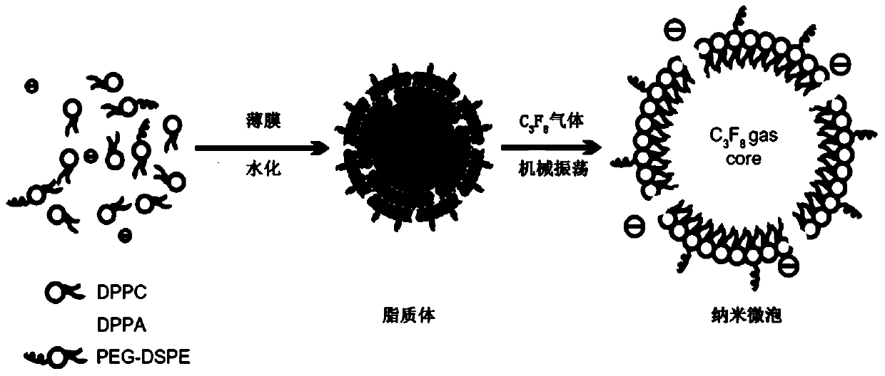
[0024]1) Dissolve 36 mg of dipalmitolecithin (DPPC), 7 mg of distearoylphosphatidylethanolamine (DSPE), and 2 mg of dipalmitophosphatidic acid (DPPA) in 8 ml of chloroform by film hydration method. Leave the chloroform to evaporate naturally in a fume hood to form a phospholipid film. Add 8 mL of hydration solution (PBS:glycerol=9:1 (volume ratio)) to the above-mentioned film-forming culture dish, and hydrate on a shaker at room temperature for 60 min. Mix the washed phospholipid film with the hydration solution and transfer it to a 50mL centrifuge tube to form liposomes (such as figure 1 shown).

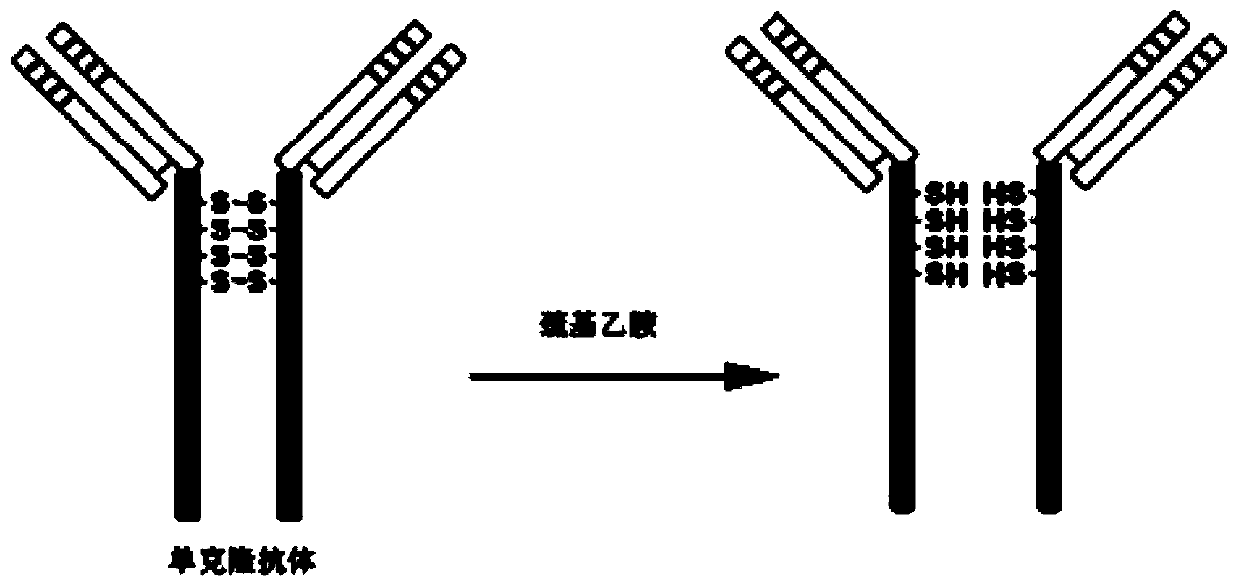
[0025] 2) Dilute 10 μg of anti-progastrin-releasing peptide monoclonal antibody to 50 μL with 10 mM PBS (Nacl 8g / L, Kcl 0.2g / L, Na2HPO4 1.44g / L), add 1 μL of 0.5M EDTA solution; Dissolve 60 mg of mercaptoethylamine in 500 μL of PBS, add 10 μL of 0.5 M EDTA solution, mix the above ...

Embodiment 2

[0027] Example 2 Identification of antibody linkages in targeted nanobubbles

[0028] Mix 100ul each of ordinary nanobubbles and targeted nanobubbles with Dylight488-labeled goat anti-mouse IgG4ul. Under a fluorescent microscope (1000×), it can be seen that the surface of targeted nanobubbles emits bright green fluorescence, while the surface of ordinary nanobubbles emits bright green fluorescence. No obvious fluorescence (such as Figure 4 shown).

Embodiment 3

[0029] Example 3 Binding experiment of targeted nano-microbubbles and small cell lung cancer (H446 cells) cells

[0030] 1.5×10 per hole 4 Cells were inoculated in a 6-well plate covered with coverslips, cultured overnight in an incubator, fixed with 4% paraformaldehyde at room temperature, added 30ul of targeted nanobubbles and normal blank nanobubbles to the slides, and kept at 37°C React in the incubator for 1 hour, observe the combination of targeted nanobubbles and cells under a microscope at 1000 times, and calculate the adhesion rate. Targeted nanobubbles adhere tightly around the H446 cells, and they are regularly arranged along the cell membrane. The average adhesion rate of the cells is (90.2±3.24)%. Figure 5 shown).


PUM


Abstract

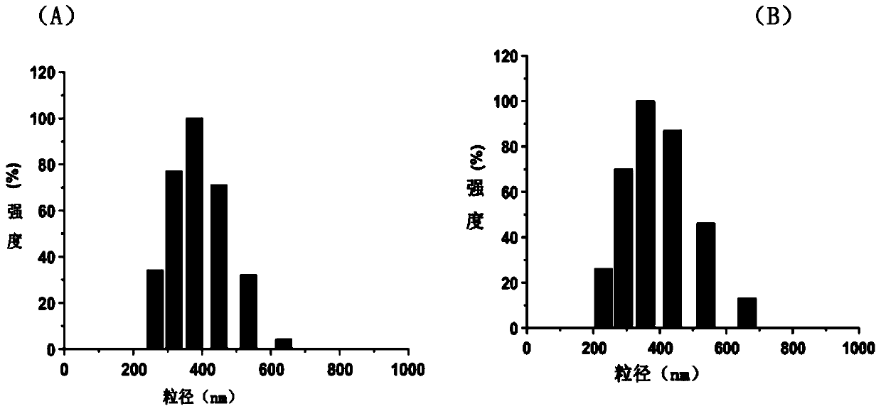
The invention provides a targeted nanobubble for detecting small cell lung cancer and a preparing method and application thereof.The targeted nanobubble comprises a lipid dimolecular shell and inert gases inside the lipid dimolecular shell, wherein an anti-progastrin releasing peptide monoclonal antibody aiming at the small cell lung cancer is connected to the lipid dimolecular shell.The average grain diameter of the nanobubbles is 300-400 nm.The preparing method comprises the steps that dipalmitoyl phosphatidylcholine, distearoyl phosphatidyl ethanolamine and dipalmitoyl phosphate are completely dissolved in chloroform to generate a lipidosome with the filming-rehydration method, the anti-progastrin releasing peptide monoclonal antibody aiming at the small cell lung cancer is opened with mercaptoethylamine for incubation with the lipidosome, so that a stable thioether bond is formed, then perfluoropropane gas is introduced, and the targeted nanobubble containing the anti-progastrin releasing peptide monoclonal antibody aiming at the small cell lung cancer is obtained.The targeted nanobubble can be applied to color doppler ultrasonic detection of small cell lung cancer.

Description

technical field [0001] The invention relates to a targeted nano-microbubble, in particular to a preparation method and application of a targeted nano-microbubble for detecting small cell lung cancer. Background technique [0002] Small cell lung cancer (small cell lung cancer, SCLC) accounts for about 20% of lung cancer, and the two-year survival rate is less than 5%. Routine tumor examinations such as X-ray, CT, and MRI lack specificity in the diagnosis of SCLC, and SCLC is often diagnosed at an advanced stage (Kalemkerian GP, ​​et a1. Small cell lung cancer. J Natl Compr Canc Netw 2013, 11(1): 78-98.). [0003] In 1981, McDonald et al. discovered that SCLC cell lines could secrete gastrin-releasing peptide (GRP) and contain GRP receptor (McDonald TJ, et al. A qualitative comparison of canine plasma gastroenteropancreatic hormone response to bombesin and the porcine gastrin-releasing peptide (GRP ). Regul Pept, 1981, 2 (5): 293-304.), but its half-life in blood is only tw...

Claims


Application Information

Patent Timeline
Patent Type & Authority Patents(China)
IPC IPC(8): A61K49/22
CPCA61K49/221A61K49/223
Inventor 王伟王金萍许静梁爱华
Owner SHANXI UNIV
Learn More