X-ray CT device, image processing device and image reconstruction method
A technology of X-ray and radiation dose, applied in the field of image reconstruction processing of projection data, which can solve problems such as image quality deviation
- Summary
- Abstract
- Description
- Claims
- Application Information
AI Technical Summary
Problems solved by technology
Method used
Image
Examples
no. 1 Embodiment approach
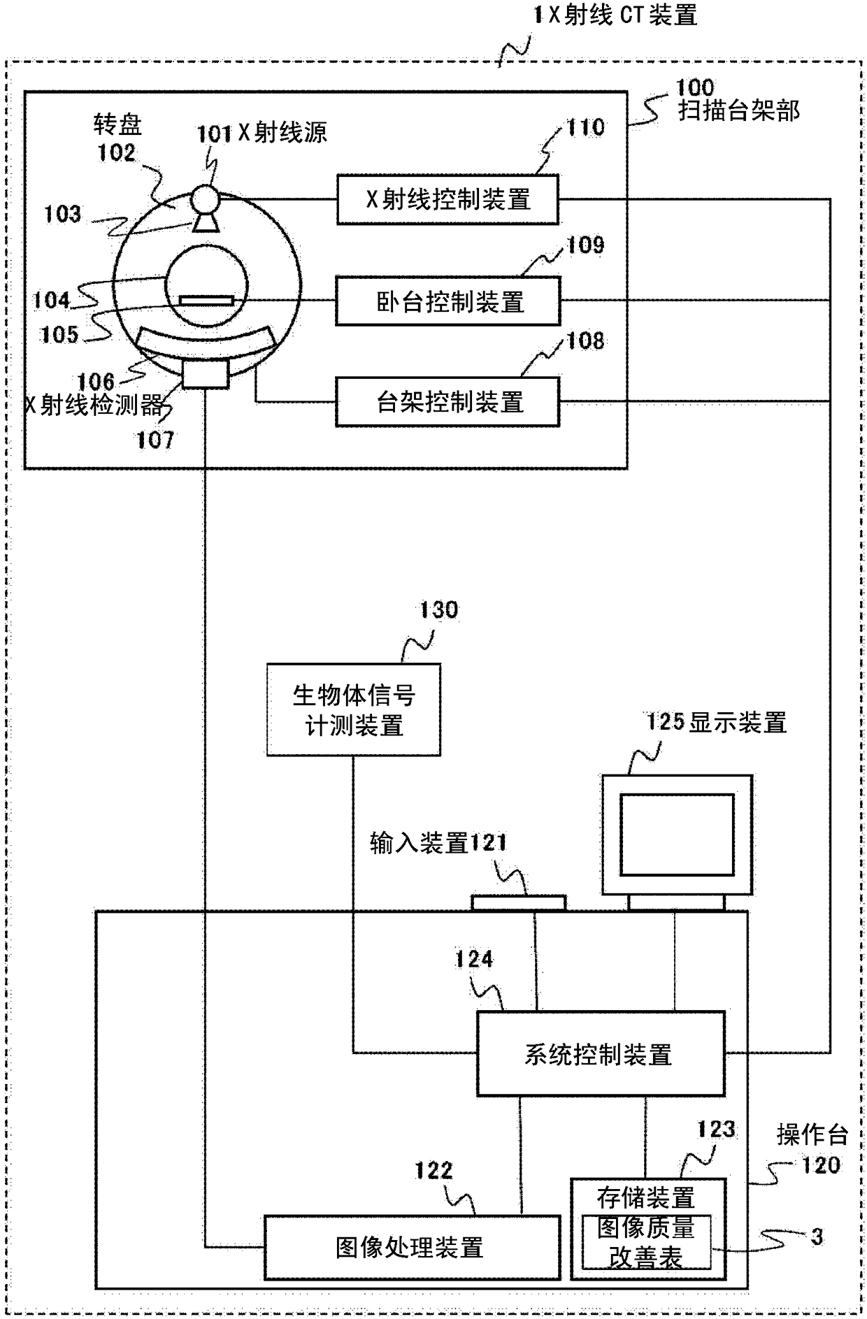
[0035] First, refer to figure 1 The overall configuration of the X-ray CT apparatus 1 will be described.
[0036] Such as figure 1 As shown, the X-ray CT apparatus 1 includes a scanning stage unit 100, a bed 105, a console 120, and a biological signal measurement device 130. The scanning gantry 100 is an apparatus that irradiates an object with X-rays and detects X-rays transmitted through the object. The console 120 is a device that controls each part of the scanning gantry 100 and acquires transmitted X-ray data measured by the scanning gantry 100 to generate an image. The bed 105 is a device that carries the subject and carries the subject in and out within the X-ray irradiation range of the scanning gantry 100. The biological signal measurement device 130 is a device that measures data related to the movement of a biological body, such as an electrocardiometer, a respirometer, and the like.
[0037] The scanning stage unit 100 includes an X-ray source 101, a turntable 102, a ...
no. 2 Embodiment approach
[0114] Next, refer to Figure 10 ~ Figure 12 The second embodiment of the present invention will be described.
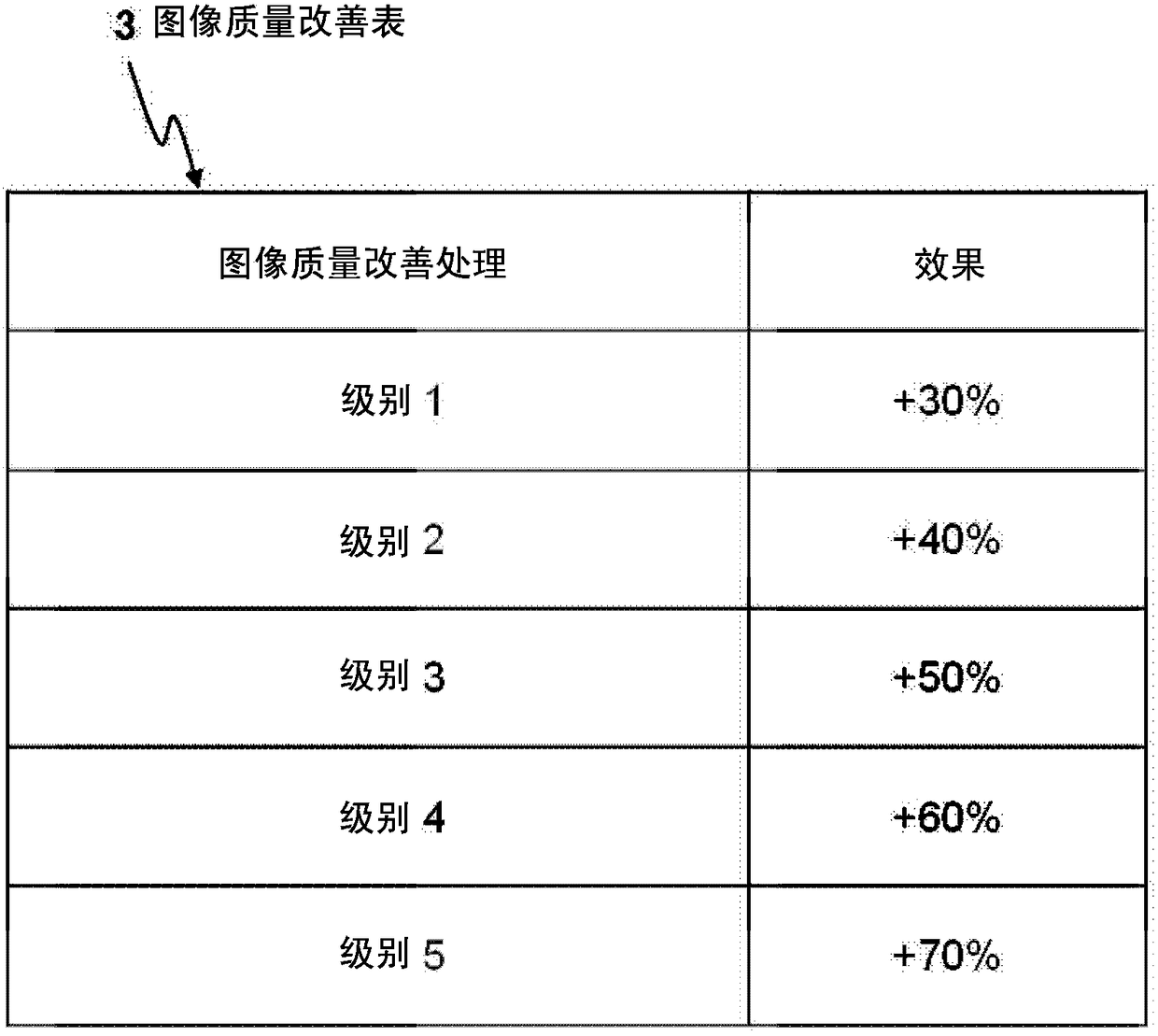
[0115] The image quality improvement effect of the image quality improvement process is limited, and there may be a relationship between the reference radiation dose and the amount of radiation at the time of shooting of the projection data that is the reconstruction target, and no matter which image quality improvement process is used. Improve to the standard image quality. For this reason, in the second embodiment, the system control device 124a of the X-ray CT apparatus 1 refers to the image quality improvement table 3 stored in the storage device 123, and obtains the lower limit value of the low dose value in advance before imaging. The ray dose modulation range setting limit.
[0116] The hardware configuration of the X-ray CT apparatus 1 of the second embodiment is figure 1 the same. In the following description, the same reference numerals are given to the same ...
no. 3 Embodiment approach
[0135] Next, refer to Figure 13 ~ Figure 15 The third embodiment of the present invention will be described.
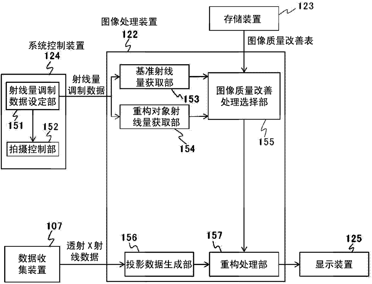
[0136] In the first embodiment, the procedure of the image quality improvement process in the image reconstruction process at the time of shooting of the subject is exemplified, but the image quality improvement process of the present invention can also be applied to projection data captured in advance and stored in the storage device 123 . In addition, in the first embodiment, the image quality as a reference is determined by the selection of the dose value or the phase, but it is also possible to display the images of each phase actually reconstructed through a moving image display or a list display, and specify them as the reference (Target) image quality.
[0137] In the third embodiment, it is also possible to describe the application of the image quality improvement process to the stored projection data and the setting of the target image quality.
[0138] The hard...
PUM
 Abstract
Description
Claims
Application Information
 - R&D
- Intellectual Property
- Life Sciences
- Materials
- Tech Scout
- Unparalleled Data Quality
- Higher Quality Content
- 60% Fewer Hallucinations
Browse by: Latest US Patents, China's latest patents, Technical Efficacy Thesaurus, Application Domain, Technology Topic, Popular Technical Reports.
© 2025 PatSnap. All rights reserved.Legal|Privacy policy|Modern Slavery Act Transparency Statement|Sitemap|About US| Contact US: help@patsnap.com


