Deep learning and MRI image-based ventricular function index prediction method

A technology of deep learning and prediction methods, which is applied to computer systems based on knowledge patterns, machine learning, character and pattern recognition, etc. It can solve problems such as high labor and time consumption, and human differences.

Inactive Publication Date: 2016-11-09
HARBIN INST OF TECH
View PDF0 Cites 36 Cited by
  • Summary
  • Abstract
  • Description
  • Claims
  • Application Information

AI Technical Summary

Problems solved by technology

[0003] The purpose of the present invention is to solve the problem that the traditional ventricular index prediction method relies on manual segmentation of the ventricular muscle part of each phase, and then calculates on this basis, and there are problems of large manpower and time consumption and serious artificial differences, and proposes a Prediction method of ventricular function index based on deep learning and MRI images

Method used


Image

  • Deep learning and MRI image-based ventricular function index prediction method
  • Deep learning and MRI image-based ventricular function index prediction method
  • Deep learning and MRI image-based ventricular function index prediction method

Examples


specific Embodiment approach 1

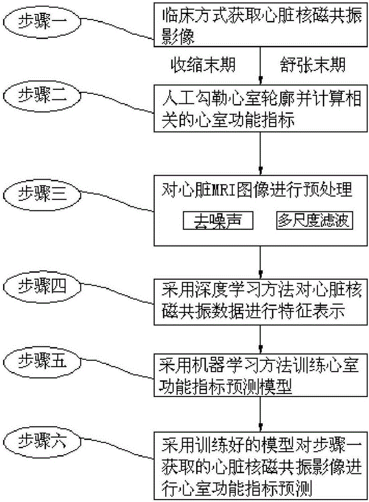
[0020] The ventricular function index prediction method based on deep learning and MRI images in this embodiment, combined with figure 1 As shown, the method is realized through the following steps:

[0021] Step 1. Obtain cardiac MRI images in a clinical manner;

[0022] Step 2, manually outline the ventricle and calculate the related ventricular function index;

[0023] Step 3, preprocessing the cardiac MRI image;

[0024] Step 4, using a deep learning method to perform feature representation on the cardiac MRI data;

[0025] Step 5, using machine learning methods to train the ventricular function index prediction model;

[0026] Step 6: Use the trained model to predict the ventricular function index on the cardiac magnetic resonance image obtained in step 1; wherein, MRI refers to nuclear magnetic resonance imaging, and is referred to as magnetic resonance imaging for short.

specific Embodiment approach 2

[0027] The difference from Embodiment 1 is that in the method for predicting ventricular function indicators based on deep learning and MRI images in this embodiment, the cardiac magnetic resonance image in step 1 is a cardiac MRI image acquired by MRI equipment, or two phases and multiple directions Among them, the two phases are the end-systole and the end-diastole, and the slice data in multiple directions are the short-axis tangential image of the base part of the cardiac MRI, the short-axis tangential image of the middle ventricle, and the short-axis tangential image of the upper ventricle , long-axis two-chamber images and long-axis four-chamber images.

specific Embodiment approach 3

[0028] The difference from Embodiment 1 or Embodiment 2 is that in the method for predicting ventricular function indexes based on deep learning and MRI images in this embodiment, the process of manually drawing the outline of the ventricle and calculating the relevant ventricular function indexes described in step 2 is as follows. The ventricular part of the acquired cardiac MRI image is manually segmented, and the ventricular function index is calculated according to the clinical general gold standard; wherein, the ventricular function index specifically includes: the volume, mass, end-systolic blood volume, and diastole of the left and right ventricles. End blood volume and ejection fraction; where mass is calculated from volume and ejection fraction is calculated from end systolic blood volume and end diastolic blood volume.

[0029] Among them, when manually segmenting the ventricle of the cardiac MRI image obtained in step 1, clinicians use general standards, and generall...


PUM


Abstract

The invention discloses a deep learning and MRI (Magnetic Resonance Imaging) image-based ventricular function index prediction method and belongs to the field of medical image processing. A conventional ventricular index prediction method mainly depends on a process of artificially segmenting a ventricular muscle part of each phase and then performing measurement and calculation based on the artificial segmentation. The process needs to consume a large amount of manpower and time and has a serious artificial difference. The deep learning and MRI image-based ventricular function index prediction method is implemented by the following steps of clinically obtaining a heart MRI image; artificially drawing a ventricular outline and calculating related ventricular function indexes; preprocessing the heart MRI image; performing characteristic representation on heart MRI data by adopting a deep learning method; training a ventricular function index prediction model by adopting a machine learning method; and performing ventricular function index prediction on the heart MRI image obtained in the first step by adopting the trained model. According to the method, the ventricular function indexes can be quickly, accurately and automatically predicted to assist in diagnosis of clinical heart diseases.

Description

technical field [0001] The invention relates to a method for predicting ventricular function indexes based on deep learning and MRI images. Background technique [0002] Medical image processing is a new discipline and technology that has developed rapidly with the development and maturity of computer technology and the advancement of clinical diagnostic technology. Nowadays, medical image processing technology is more and more widely used in clinical practice. Heart disease is one of the diseases with the highest fatality rate, so it has been paid more and more attention in clinical diagnosis and treatment. At the same time, the related technologies of heart disease detection and treatment have also become hot spots and difficulties in the research and development of medical image processing. Ventricular disease is a common heart disease, such as ventricular muscle hypertrophy, ventricular fibrillation, heart failure, etc. Due to the high soft tissue resolution capability ...

Claims


Application Information

Patent Timeline
Patent Type & Authority Applications(China)
IPC IPC(8): G06K9/62G06N5/00
CPCG06N20/00G06V2201/031G06F18/24
Inventor 王宽全骆功宁安然董素宇张恒贵
Owner HARBIN INST OF TECH
Why Eureka
Try Eureka