Target-specific dose & scatter estimation in CT images

一种投影图像、散射图像的技术,应用在估计CT图像中的散射效应领域,能够解决不准确测量等问题

Active Publication Date: 2016-11-23
ELEKTA AB +1
View PDF4 Cites 11 Cited by
  • Summary
  • Abstract
  • Description
  • Claims
  • Application Information

AI Technical Summary

Problems solved by technology
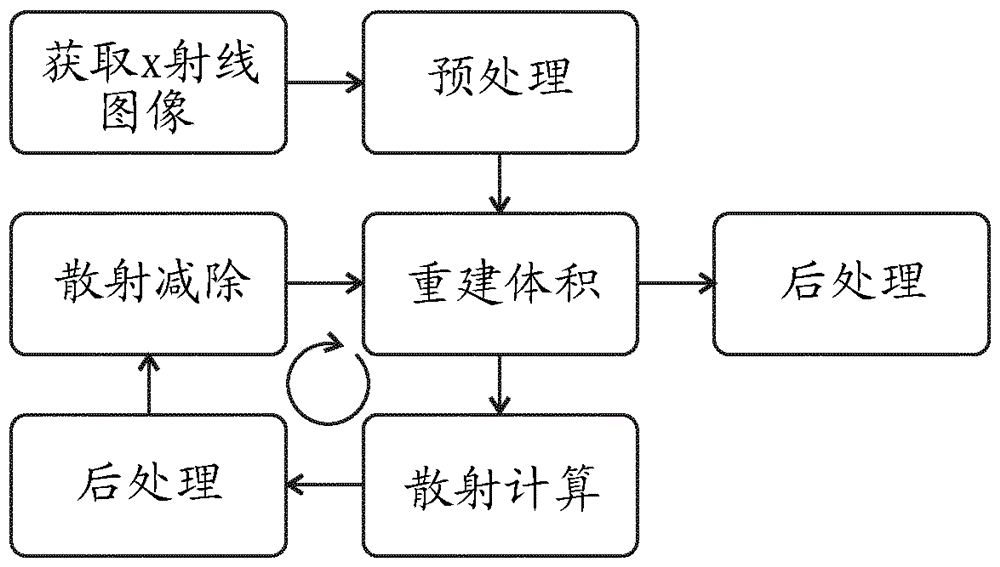
In the latter case, photons are re-emitted in random directions and thus can be detected, leading to inaccurate measurements of attenuation elsewhere in the projected image

Method used


Image

  • Target-specific dose & scatter estimation in CT images
  • Target-specific dose & scatter estimation in CT images
  • Target-specific dose & scatter estimation in CT images

Examples


Embodiment Construction

[0031] refer to figure 1 , shows a conventional radiotherapy setup. This comprises a vertically arranged gantry 10 rotatable about a transverse axis. A patient support 12 is placed just below this axis and carries a patient 14 . The patient support 12 is generally adjustable with sufficient degrees of freedom to position the patient as desired so that the target area of ​​the patient is correctly positioned relative to the device. A radiotherapy source 16 is mounted on the gantry 10 and emits a beam of therapeutic radiation toward the patient 14, typically having a photon energy of 1 MeV or above. Such radiation is harmful to tissue and, if properly directed, can have a therapeutic effect on lesions such as tumors in a patient. To maximize the radiation's effect on the lesion and minimize its effect on surrounding healthy tissue, the beam is aimed with a collimator within the source 16, and the source itself is gantry rotated around the patient. The dose rate of the beam, ...


PUM


Abstract

Scatter correction of the projection images used to reconstruct volume CT images is a computationally intensive process that cannot at present be done in real time. We note, however, that although scatter is non-uniform, it varies smoothly - i.e. with a low spatial frequency. Existing Monte-Carlo estimation methods need to run over a large number of iterations in order to eliminate the mainly high-frequency artefacts that are present initially, but this process can be cut short and a low-pass filter applied instead thereby substantially reducing the computational load. By applying the same principle in the spatial domain and interpolating some scatter images from adjacent and surrounding scatter images, the computational load can be reduced still further. Applying these techniques in an iterative process on graphics-processor-quality computational hardware enables real-time scatter correction and reconstruction of CT volume images.

Description

technical field [0001] The invention relates to a method of estimating the effect of scatter in CT images and a device embodying the method. This can be used to prepare images in which the effects of this scattering are mitigated. Background technique [0002] A CT reconstruction is a multi-dimensional image showing the internal structure of a patient or object. It is prepared by taking many x-ray projections through the patient or object, which are then used in the calculation process to calculate the structure that would have resulted in the set of x-ray projections used. [0003] Various forms of x-ray projections are available. A single narrow beam (or "pencil beam") can be used along which the attenuation of the x-ray beam will be measured. It is then rotated around the patient or object, measuring attenuation along a range of directions. This allows determination of the internal structure of a single "slice", which is the plane in which the beam rotates. More gene...

Claims


Application Information

Patent Timeline
Patent Type & Authority Applications(China)
IPC IPC(8): A61B6/03A61B6/00G06T11/00G06T5/00
CPCA61B6/032A61B6/5205A61B6/5282G06T5/50G06T2207/10081G06T2207/20076G06T2207/20224G06T2207/30004G06T11/005G06T11/003A61B6/4014G06T5/70G01N23/046G06T2207/10116G06T5/00G06T7/10G06F18/22G06T7/0012G06T7/20G06T11/008G06T2207/20112G06T2207/20182
Inventor M.亨尼克斯H.诺德斯特伦M.埃里克森J.阿德勒D.贾弗雷G.布茨马F.弗赫根B.努蒂
Owner ELEKTA AB
Why Eureka
Try Eureka