

Automatic retinal vessel segmentation method based on non-fluorescent fundus images
A technology for automatic segmentation of retinal blood vessels, which is applied in the field of image processing, can solve the problems of low segmentation efficiency, low segmentation accuracy of tiny blood vessels, and easy adhesion, etc., and achieve the effects of suppressing uneven illumination, improving segmentation effects, and increasing contrast
- Summary
- Abstract
- Description
- Claims
- Application Information
AI Technical Summary
Problems solved by technology
Method used
Image

Examples
Embodiment
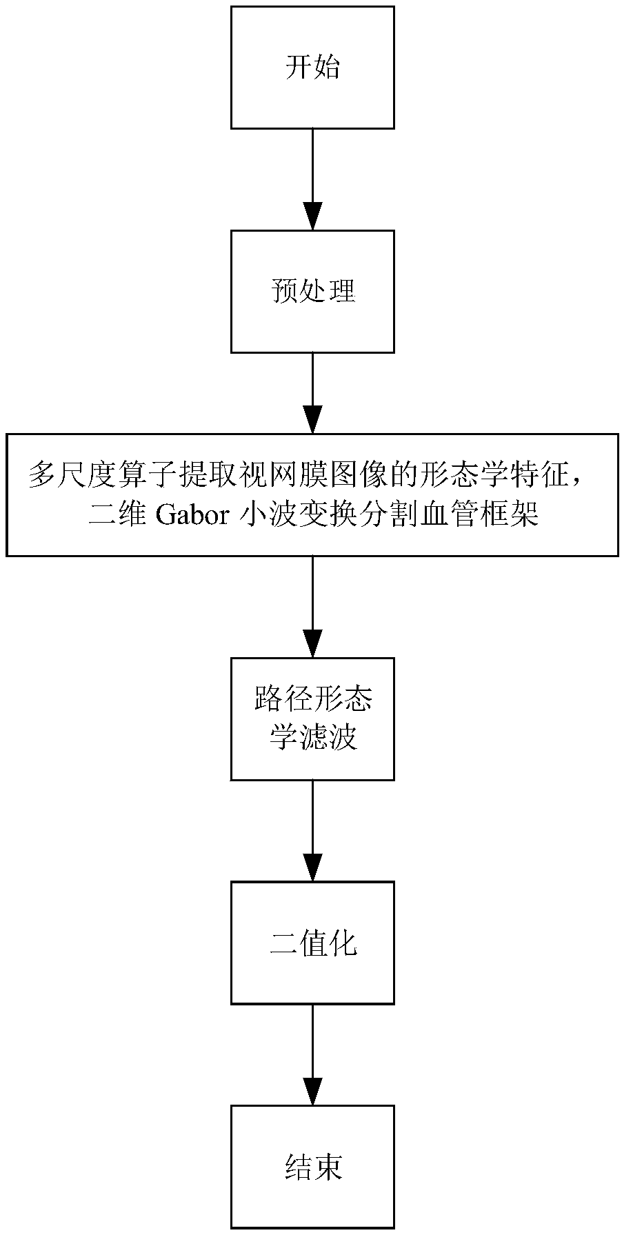
[0047] This embodiment includes the following steps:
[0048] The first step is to preprocess the image to enhance the characteristics of blood vessels and weaken the background noise.
[0049] The preprocessing includes contrast enhancement and retinal border growth.
[0050] Contrast enhancement is mainly based on a contrast-limited adaptive histogram equalization algorithm. The R channel is overexposed and the contrast is low; the brightness of the B channel is low, and blood vessels are difficult to identify; compared with the R and B channels, the contrast between the blood vessels and the background of the G channel image is the highest, and the noise is less. Therefore, we choose the G channel image for subsequent processing. The present invention uses the CLAHE algorithm to improve the local contrast of the G channel image, expecting to present more image details. Compared with the common adaptive histogram equalization method, the characteristic of CLAHE lies in it...
PUM
 Abstract
Description
Claims
Application Information