Fluorescent probe with charge transfer properties and preparation method and use thereof

A fluorescent probe, charge transfer technology, applied in fluorescence/phosphorescence, chemical instruments and methods, organic chemistry, etc., can solve problems such as the need to improve the selectivity of action, and achieve low preparation cost, large market competitiveness, and probe structure. stable effect

Active Publication Date: 2019-04-02
ANHUI IPROCOM BIOTECH CO LTD
View PDF3 Cites 0 Cited by
  • Summary
  • Abstract
  • Description
  • Claims
  • Application Information

AI Technical Summary

Problems solved by technology

Many small molecules that recognize G-quadruplexes have a certain binding ability to double-helix DNA, so the selectivity of the interaction between small molecules and G-quadruplex structures needs to be improved

Method used


Image

  • Fluorescent probe with charge transfer properties and preparation method and use thereof
  • Fluorescent probe with charge transfer properties and preparation method and use thereof
  • Fluorescent probe with charge transfer properties and preparation method and use thereof

Examples


Embodiment 1

[0039] Example 1: Synthesis of Fluorescent Probes

[0040] Dissolve 0.27 g of 4-formyltriphenylamine in 20 mL of n-butanol, add 0.38 g of 4-chloro-1,2-dimethylquinoline iodonium and 0.11 g of N,N-dimethylethyl Diamine, reacted at 120°C for 15h. After the reaction, it was naturally cooled to room temperature, and the solvent was distilled off. Add 50 mL of dichloromethane and stir for 10 minutes, filter to obtain a solid product, and then wash the solid twice with 20 mL of dichloromethane to obtain 0.21 g of pure product. The yield was 34.4%. H NMR 1 H NMR (400MHz, DMSO-d 6 )δ: 9.45(s, 1H), 8.86(s, 1H), 8.49(d, J=8.12Hz, 1H), 8.26(d, J=8.84Hz, 1H), 8.07(t, J=7.52Hz, 1H), 7.84-7.78(m, 4H), 7.56(d, J=15.76Hz, 1H), 7.40-7.36(m, 4H), 7.17-7.10(m, 6H), 6.99(d, J=8.12Hz ,2H),4.16(s,3H),4.03(s,2H),3.46(s,2H),2.90(s,6H). C NMR 1 C NMR (100MHz, DMSO-d 6 )δ:155.45,154.19,149.84,146.91,142.77,139.96,134.56,130.54,130.43,128.90,126.85,125.74,124.92,121.56,119.57,118.32,118.08,97.98...

Embodiment 2

[0041] Embodiment two: the stability of fluorescent probe

[0042] 1. The fluorescent probe was prepared into a 5 mM stock solution with DMSO solvent, and then diluted with a 10 mM Tris-HCl buffer solution (pH 7.4, containing 60 mM KCl) to a 10 μM probe solution for testing.

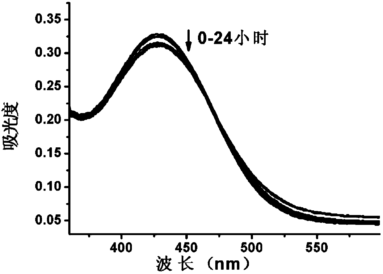
[0043] 2. Determination of ultraviolet absorption spectrum. Test the absorption spectrum of the above-mentioned probe solution at 360-600nm at the time of 0, 0.1, 0.5, 1, 2, 5, 7, 11, 20, and 24 hours with a UV spectrophotometer, so as to determine the fluorescence of the fluorescent probe. stability. The result is as figure 1 As shown, the ultraviolet absorption of the probe did not change significantly within 0-24 hours, and no new absorption peak appeared, indicating that the probe could stably exist in the solution under the experimental conditions.

Embodiment 3

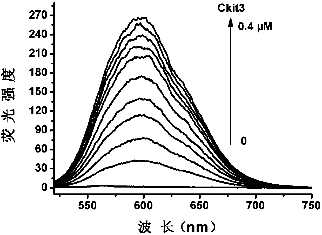
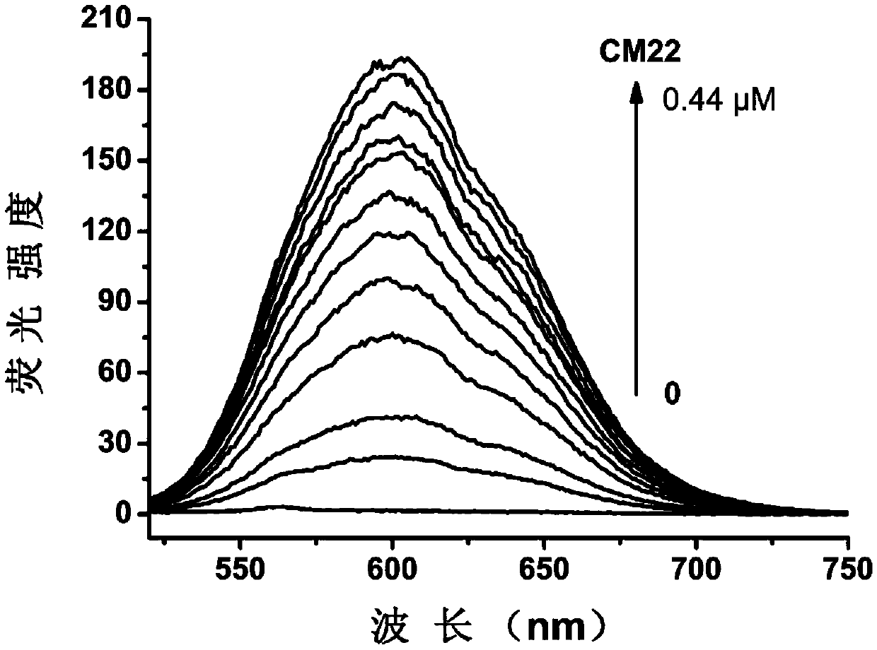
[0044] Embodiment three: the detection of DNA sample

[0045] 1. DNA sample preparation. DNA samples were purchased from Shanghai Sangon Co., Ltd. The DNA sample was dissolved in an appropriate amount of buffer solution (10mM Tris-HCl, pH 7.4, 60mM KCl), and its concentration was calculated according to the ultraviolet absorption value and molar absorptivity of the sample solution at 260nm.

[0046] Wherein, the DNA sequence used is:

[0047] Ckit3: GGCGAGGAGGGGCGTGGCCGGC

[0048] CM22: TGAGGGTGGGTAGGGTGGGTAA

[0049] HRAS: TCGGGTTGCGGGCGCAGGGCACGGGCG

[0050] c-myc: TTGAGGGTGGGTAGGGTGGGTAAA

[0051] 22AG: AGGGTTAGGGTTAGGGTTAGGG

[0052] G3T3: GGGTTTGGGTTTGGGTTTGGG

[0053] htg-21: GGGTTAGGGTTAGGGTTAGGG

[0054] Ckit1: AGGGAGGGCGCTGGGAGGAGGG

[0055] ds26: CAATCGGATCGAATTCGATCCGATTG

[0056] polyd(A-T) 9 : ATATATATATATATATAT

[0057] polyd (G-C) 9 : GCGCGCGCGCGCGCGCGC

[0058] ct-DNA: calf thymus DNA

[0059] ss26: CCGCGAACGCCTAAGCTGCTAACCGC

[0060] 2. Prepara...


PUM


Abstract

The invention discloses a fluorescent probe with the characteristic of charge transfer, and a preparation method and an application thereof, belonging to the fields of chemical synthesis, chemical analysis and biological analysis. According to the invention, the structure of the probe comprises the parts of electron-donating triphenylamine and electron-deficient quinoline salt, so the probe has the property of intramolecular charge transfer. The fluorescent probe has the following advantages: the synthetic route has high operability; the purification method is simple; the preparation cost is low; and good market competitiveness is obtained. The fluorescent probe has stable structure and low fluorescence background, wherein the fluorescence enhancement exceeds one hundred times after specific binding of the fluorescent probe with a G-quadruplex. Through fluorescence spectrum, a secondary structure of a DNA sample can be rapidly detected, and the disadvantages like high price, long cycle and complicated technical operation in the prior art are overcome.

Description

technical field [0001] The invention relates to the construction of a novel fluorescent probe, its preparation method and application, and belongs to the fields of chemical synthesis, chemical analysis and biological analysis. Background technique [0002] G-quadruplex DNA is a special DNA structure. It is a single-stranded DNA sequence rich in guanine bases. Under certain conditions, its four guanine rings self-assemble into a G-tetrad through Hoogsteen hydrogen bonds. An advanced DNA secondary structure formed by two or more G-tetrads through π-π stacking. In recent years, bioinformatics studies have shown that there are about 370,000 groups of guanine-rich gene sequences that may form a G-quadruplex structure in the human body, especially at the ends of human telomeres and oncogene promoter regions (such as c -myc, ckit, bcl-2, Pu27, kRAS, VEGFR, TERT, etc.) are more common. The G-quadruplex structure plays an important role in maintaining chromosome stability and the e...

Claims


Application Information

Patent Timeline
Patent Type & Authority Patents(China)
IPC IPC(8): C07D215/46C09K11/06G01N21/64
CPCC07D215/46C09K11/06C09K2211/1029G01N21/6428
Inventor 王明齐唐彩平刘硕刘瑞江孙竞
Owner ANHUI IPROCOM BIOTECH CO LTD
Why Eureka
Try Eureka