
Method and system for segmenting optic cup in fundus photography

An optic cup and optic disc technology, applied in the field of medical image processing, can solve the problems of easily missing local features of the optic cup, not making full use of blood vessel information, affecting the accuracy of glaucoma diagnosis results, and achieving the effects of reducing labor and improving accuracy.

Active Publication Date: 2019-09-13
BEIJING UNIV OF CHEM TECH
View PDF3 Cites 0 Cited by
  • Summary
  • Abstract
  • Description
  • Claims
  • Application Information

AI Technical Summary

Problems solved by technology

These two methods ultimately use ellipses to fit the optic cup boundary, and neither fully utilizes blood vessel information, so it is easy to miss the local features of the optic cup, thus affecting the accuracy of glaucoma diagnosis results

Method used


Image

  • Method and system for segmenting optic cup in fundus photography
  • Method and system for segmenting optic cup in fundus photography
  • Method and system for segmenting optic cup in fundus photography

Examples


Embodiment Construction

[0025] In order to make the purpose, technical solutions and advantages of the embodiments of the present invention clearer, the technical solutions in the embodiments of the present invention will be clearly and completely described below in conjunction with the drawings in the embodiments of the present invention. Obviously, the described embodiments It is a part of embodiments of the present invention, but not all embodiments. Based on the embodiments of the present invention, all other embodiments obtained by persons of ordinary skill in the art without creative efforts fall within the protection scope of the present invention.

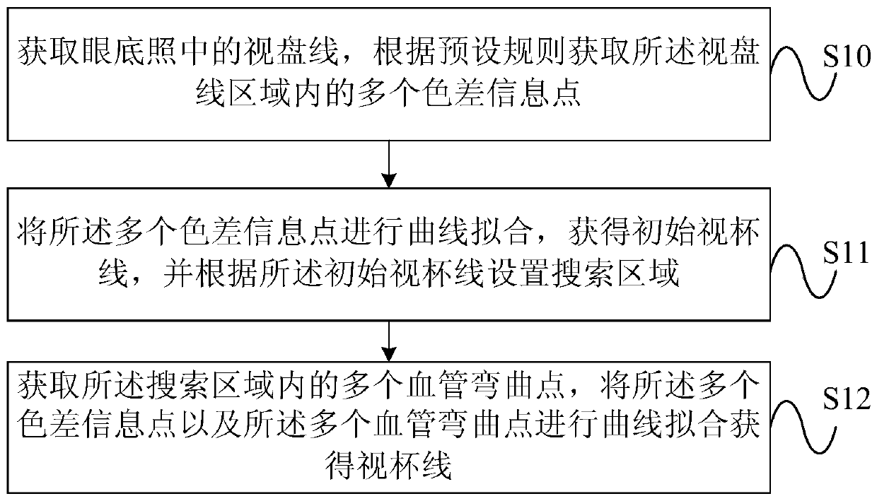
[0026] figure 1 It is a schematic flow chart of the method for segmenting the optic cup according to the fundus in the embodiment of the present invention, as figure 1 As shown, the present embodiment discloses a method for segmenting the optic cup in fundus photography, including:

[0027] S10. Obtain the optic disc line in the fundus photo, an...


PUM


Abstract

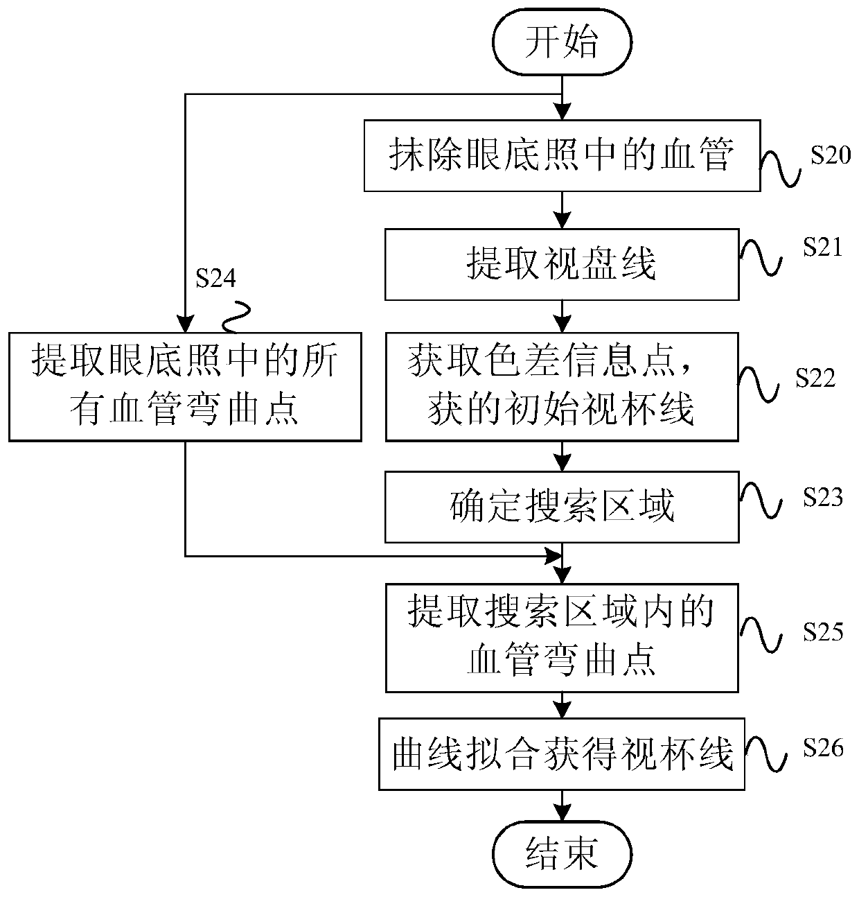
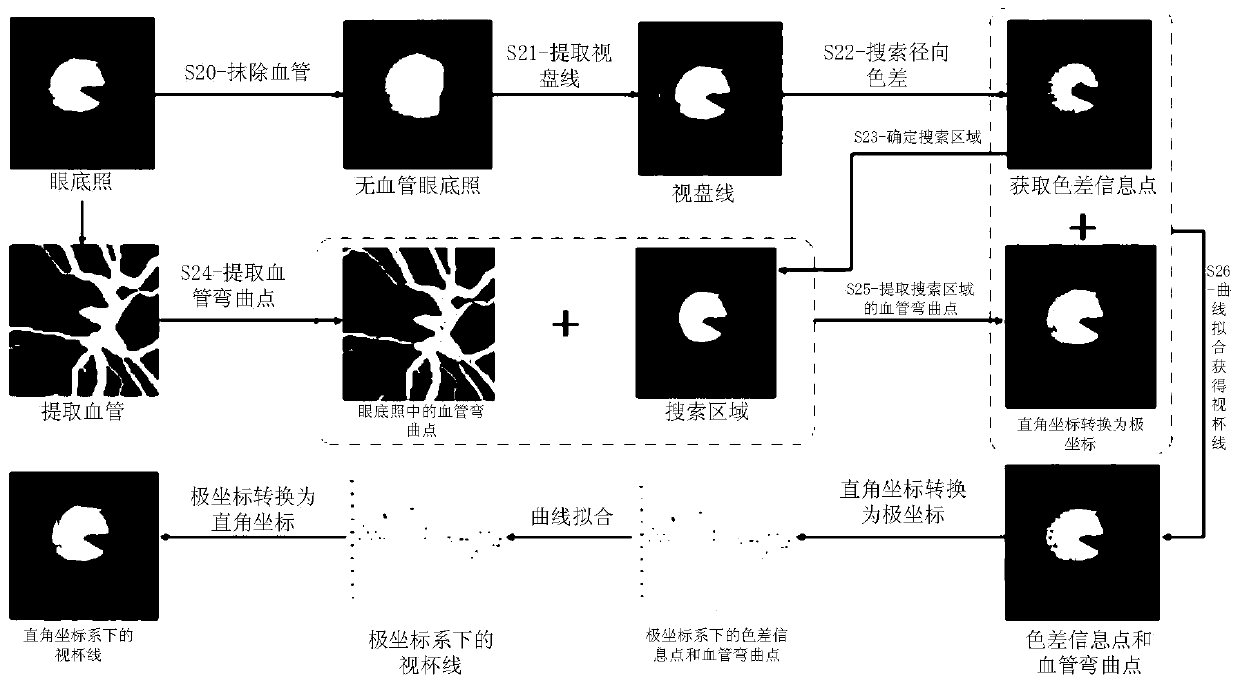
Embodiments of the present invention provide a method and system for segmenting an optic cup in a fundus photograph. The method includes: acquiring the optic disc line in the fundus photo, acquiring a plurality of color difference information points in the region of the optic disc line according to preset rules; performing curve fitting on the plurality of color difference information points to obtain the initial optic disc line, And setting a search area according to the initial optic cup line; obtaining a plurality of vascular bending points in the search area, performing curve fitting on the plurality of color difference information points and the plurality of vascular bending points to obtain an optic cup line. The system is used to perform the method described above. The method and system for segmenting the optic cup in the fundus photo provided by the embodiments of the present invention comprehensively utilize the color difference information and blood vessel information in the fundus photo to improve the accuracy of the cup segmentation and further improve the accuracy of glaucoma diagnosis results.

Description

technical field [0001] Embodiments of the present invention relate to the technical field of medical image processing, and in particular to a method and system for segmenting an optic cup in fundus photography. Background technique [0002] Eyes are one of the most important organs of the human body. If you suffer from eye diseases, your quality of life will be greatly affected. Glaucoma is a kind of eye disease. It is a progressive optic neuropathy characterized by the structural changes of the optic disc (also known as the optic nerve head). Optic nerve cells are degenerating. Since the lost function of the optic nerve can no longer be restored, early detection and timely treatment are a key to saving the vision of glaucoma patients. [0003] Fundus photography is an important examination for ophthalmologists to diagnose glaucoma. Fundus photography refers to the photos obtained by taking pictures of the fundus with fundus photography technology. Fundus photography tech...

Claims


Application Information

Patent Timeline
Patent Type & Authority Patents(China)
IPC IPC(8): G06K9/00G06T7/10
CPCG06T2207/30041G06V40/18
Inventor 徐永利
Owner BEIJING UNIV OF CHEM TECH
Learn More