A digital X-ray image stitching method and system

An image stitching and X-ray technology, applied in image enhancement, image analysis, medical images, etc., can solve the problems of patients receiving a large radiation dose, high input images, wrong stitching, etc., to improve the accuracy of stitching and reduce radiation dose. , the effect of overcoming splicing errors

Active Publication Date: 2020-01-07
深圳蓝影医学科技股份有限公司
View PDF5 Cites 0 Cited by
  • Summary
  • Abstract
  • Description
  • Claims
  • Application Information

AI Technical Summary

Problems solved by technology

This method has relatively high requirements on the input image, and it is prone to false splicing; this method requires multiple exposures, and the patient needs to receive a large radiation dose

Method used


Image

  • A digital X-ray image stitching method and system
  • A digital X-ray image stitching method and system
  • A digital X-ray image stitching method and system

Examples


Embodiment

[0036] The present invention proposes a digital X-ray image splicing method and system, which only requires the doctor to simply select the shooting range of the patient, and the system will automatically complete the process of splicing images with only one exposure in a short time. This process does not require human intervention, the system operates reliably and the image stitching effect is good.

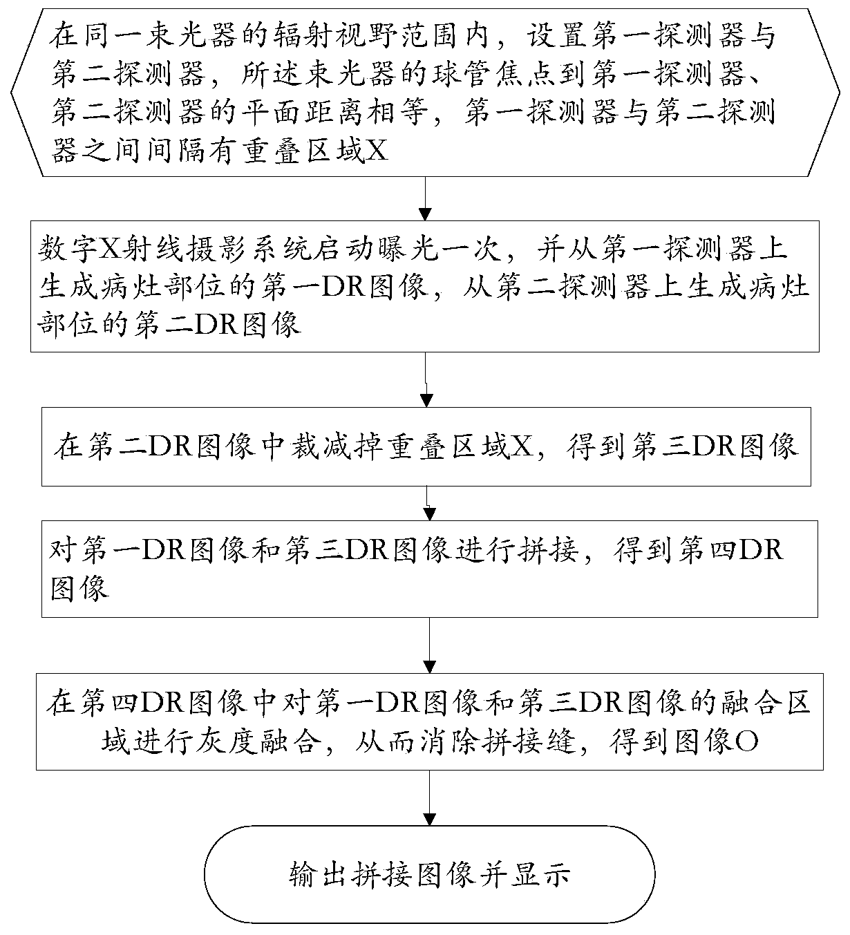
[0037] Such as figure 1 , a digital X-ray image stitching method of the present invention, comprises the following steps:

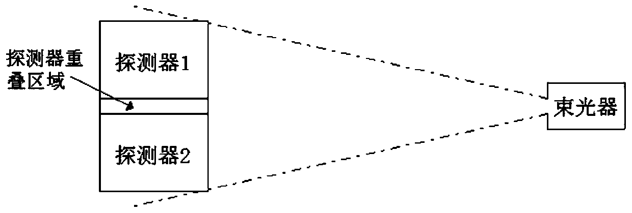
[0038] Step A. Set the first detector and the second detector within the radiation field of view of the same light beam device. The plane distances from the focal point of the tube of the light beam device to the first detector and the second detector are equal. There is an overlapping area X between the first detector and the second detector;

[0039] Step B. The digital X-ray imaging system starts exposure once, and generates a first DR image of the les...


PUM


Abstract

The invention discloses a digital X-ray image splicing method and system. The digital X-ray image splicing method comprises steps that a first detector and a second detector are arranged in a radiation vision field scope of a same beamer, plane distances from a bulb focus of the beamer to the first detector and the second detector are identical, an overlapping region X is arranged between the first detector and the second detector with intervals; a digital X-ray shooting system starts for exposure for one time, a first DR image of a focus portion is generated on the first detector, and a second DR image of the focus portion is generated on the second detector; the overlapping region X is cut from the second DR image to acquire a third DR image; the first DR image and the third DR image are spliced to acquire a fourth DR image; gray fusion for the splicing region is carried out to eliminate a splicing seam to acquire an image O; the image O is displayed. The method is advantaged in that the spliced image can be acquired through one-time exposure, and thereby a radiation dose and the patient positioning time can be greatly reduced.

Description

technical field [0001] The present invention relates to the field of digital image processing, in particular the present invention relates to digital image processing applied to medical purposes, in particular to a digital X-ray image splicing method and system. Background technique [0002] With the continuous development of medical technology and medical diagnosis methods, digital X-ray images have been widely used in the fields of clinical diagnosis and medical research, especially in orthopedic diagnosis and treatment, which has very important clinical application value. In some clinical diagnosis and surgical treatment, images of the whole body of the patient are required for comprehensive and intuitive observation and analysis of the lesion and its surroundings. Currently limited by the size of the detector, digital X-ray imaging systems commonly used in orthopedic clinics can only provide images of parts of the patient's body. Clinical diagnosticians need to analyze a...

Claims


Application Information

Patent Timeline
Patent Type & Authority Patents(China)
IPC IPC(8): G06T3/40G06T7/10G06T5/00A61B6/00G16H30/20
CPCA61B6/5241G06F19/321G06T3/4038G06T5/005G06T2207/10124G06T2207/20132G06T2207/30008
Inventor 郭朋郑杰李勇
Owner 深圳蓝影医学科技股份有限公司
Why Eureka
Try Eureka