Image processing method and device of anal sphincter and ultrasonic device

An anal sphincter and image processing technology, which is applied in image data processing, image enhancement, image analysis, etc., can solve problems such as cumbersome operation, unfavorable user identification, and inability to guarantee the accuracy of manual measurement

Active Publication Date: 2017-05-17
SONOSCAPE MEDICAL CORP
View PDF5 Cites 3 Cited by
  • Summary
  • Abstract
  • Description
  • Claims
  • Application Information

AI Technical Summary

Problems solved by technology

Due to the small area of ​​each image, it is not only not conducive to user identification, but also cannot guarantee the accuracy of manual measurement
If the identification and me

Method used


Image

  • Image processing method and device of anal sphincter and ultrasonic device
  • Image processing method and device of anal sphincter and ultrasonic device
  • Image processing method and device of anal sphincter and ultrasonic device

Examples


Embodiment Construction

[0046] The embodiments of the present invention will be further described in detail below in conjunction with the drawings and embodiments. It should be understood that the specific embodiments described here are only used to explain the embodiments of the present invention, rather than to limit the embodiments of the present invention. In addition, it should be noted that, for the convenience of description, the drawings only show some but not all structures related to the embodiments of the present invention.

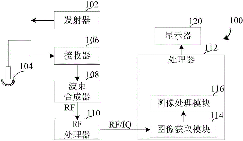
[0047] figure 1 It is a structural block diagram of an ultrasonic device 100 according to an embodiment of the present invention. The system 100 includes a transmitter 102. A probe 104 transmits an ultrasonic signal to an object according to a driving signal applied from the ultrasonic transmitter 102. A receiver 106 receives ultrasonic echoes reflected from the object. Signal. The probe 104 may include a plurality of transducers that vibrate according to electrical...


PUM


Abstract

Disclosed are an image processing method and a device of a anal sphincter and an ultrasonic device. The image processing method of the anal sphincter comprises obtaining 3D volume data of an inspector when in an anal contraction state, obtaining a multi-frame section plane image by a multi-plane imaging of the 3D volume data, and identifying the region of the internal anal sphincter and the region of the external anal sphincter in each frame according to a sonographic feature of the anal sphincter. The image processing method and a device of the anal sphincter and an ultrasonic device can recognize the form of the internal and external anal sphincter quickly and accurately.

Description

technical field [0001] Embodiments of the present invention relate to the technical field of ultrasound equipment, and in particular to an anal sphincter image processing method and device, and ultrasound equipment. Background technique [0002] Three-dimensional ultrasonography has the advantages of good repeatability, no radiation, and low cost, and is currently an effective way to evaluate pelvic floor function. To detect anal sphincter injury on multiple planes of three-dimensional ultrasound, a multi-frame arrangement is generally adopted, and the imaging range is from above the external anal sphincter to below the internal anal sphincter. Due to the small area of ​​each image, it is not only not conducive to user identification, but also cannot guarantee the accuracy of manual measurement. If the identification and measurement are carried out in a single or enlarged state on multiple planes, it is necessary to perform multiple state switching on multiple images, which...

Claims


Application Information

Patent Timeline
IPC IPC(8): G06T7/00G06T7/11G06T7/136
CPCG06T7/0012G06T2207/10136G06T2207/20004G06T2207/30004
Inventor 李萍唐艳红党静田秋芳许龙
Owner SONOSCAPE MEDICAL CORP
Why Eureka
Try Eureka