
Microfluidic chip for detection of tumor marker group

A tumor marker and microfluidic chip technology, which is applied in the field of microfluidic chips for tumor marker group detection, can solve problems such as restricting the promotion of tumor screening work and being difficult to benefit low-income groups.

Active Publication Date: 2017-05-24
孙翠敏
View PDF7 Cites 7 Cited by
  • Summary
  • Abstract
  • Description
  • Claims
  • Application Information

AI Technical Summary

Problems solved by technology

[0003] At present, tumor screening generally uses low-dose CT scanning or serum tumor marker detection, both of which need to be carried out in medical institutions with a certain scale. Widely carried out
High prices are also difficult to benefit low-income groups, which restricts the promotion of tumor screening

Method used


Image

  • Microfluidic chip for detection of tumor marker group
  • Microfluidic chip for detection of tumor marker group
  • Microfluidic chip for detection of tumor marker group

Examples


Embodiment Construction

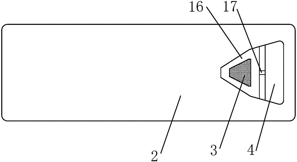
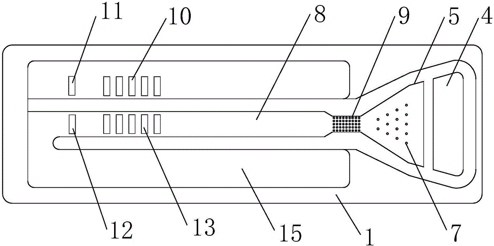
[0015] Such as figure 1 , 2 , 3, a microfluidic chip for tumor marker group detection, including a substrate 1 and a cover plate 2 above it, the cover plate 2 is made of a transparent material, and a blood collection microneedle array is arranged on the cover plate 2 3. The blood collection microneedle array 3 is composed of a plurality of blood collection microneedles, and each blood collection microneedle has a microtube 14 inside; the cover plate 2 and the base plate 1 are glued or thermally bonded to form an overflow cavity 4 , sampling chamber 16, reaction chamber 5, capillary flow channel 8 and waste liquid chamber 15, described sampling chamber 16 and reaction chamber 5 are communicated on the vertical space, sampling chamber 16 is on, and reaction chamber 5 is below; Absorbent paper 6 and filter paper 18 are arranged in the sample injection chamber 16; the capillary flow channel 8 communicates with the reaction chamber 5 through the delay valve 9; the bottom of the ta...


PUM


Abstract

The invention relates to a microfluidic chip for detection of a tumor marker group; the microfluidic chip comprises a base plate and a cover plate positioned above the base plate; the cover plate is provided with a blood collecting microneedle array, and the interior of each blood collecting microneedle is provided with a microtube; the cover plate and the base plate are glued or are subjected to thermocompression bonding to form an overflow chamber, a sample introduction chamber, a reaction chamber, a capillary flow passage and a waste liquid chamber; the sample introduction chamber communicates with the reaction chamber in a vertical space; the sample introduction chamber is internally provided with water absorbent paper and filter paper; the reaction chamber is located below the filter paper, the reaction chamber is internally provided with a microcolumn array, the bottom surface of the reaction chamber and the surface of the microcolumn array are coated with labelled antibodies coupled with chromophoric groups; the bottom of a tail end region of the capillary flow channel is provided with a plurality of test strips and quality control strips printed with immobilized antibody reagents. The self-service detection is achieved, assistance of professional devices is not needed, and an examinee operates the device by self. The blood collecting volume is extremely low, and only a drop of fingertip blood is needed and can be used for detecting various tumor marker indexes at the same time.

Description

technical field [0001] The invention relates to the technical field of microfluidics and tumor screening, in particular to a microfluidic chip for detecting tumor marker groups. Background technique [0002] The incidence of malignant tumors in our country is increasing day by day, seriously threatening people's health. The best countermeasure to deal with the threat of tumor is "early detection and early treatment". "Early detection" is the premise of "early treatment", which will greatly improve the cure rate and save a lot of medical expenses for patients and society. Tumor screening is an important way to detect cancer and precancerous lesions in the early stage. The popularization and popularization of tumor screening and the improvement of early diagnosis rate are the main effective means of diagnosis and treatment of malignant tumors. [0003] At present, tumor screening generally uses low-dose CT scanning or serum tumor marker detection, both of which need to be ...

Claims


Application Information

Patent Timeline
IPC IPC(8): G01N33/574G01N33/558
CPCG01N33/558G01N33/57484
Inventor 尤晖孙翠敏尤翔
Owner 孙翠敏
Learn More