Nano material for tumor imaging and treatment and preparation method of nano material
A nanomaterial and tumor imaging technology, applied in the field of nanomaterials, can solve problems such as large side effects and poor targeting effect of tumor treatment drugs, and achieve the effects of less toxic side effects, low equipment requirements, and easy operation.
- Summary
- Abstract
- Description
- Claims
- Application Information
AI Technical Summary
Problems solved by technology
Method used
Image

Examples
Embodiment 1
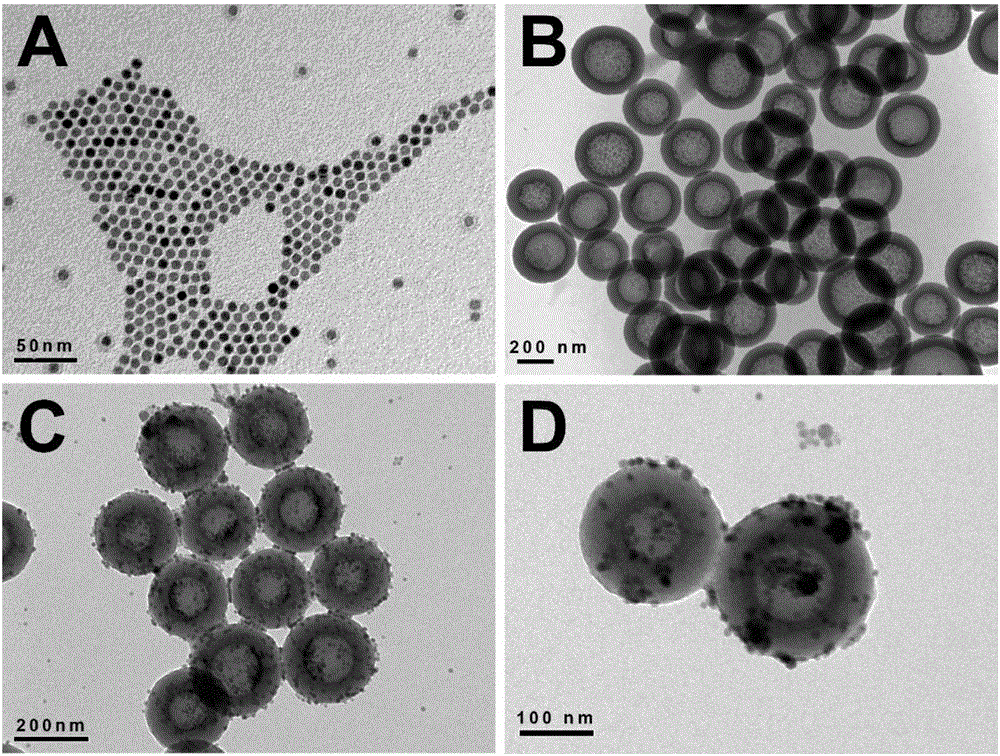
[0037] The embodiment of the present invention provides a nano material for tumor imaging and treatment and a preparation method thereof, including the following steps:
[0038] (1) Add FeCl 3 ·6H 2 O and sodium oleate (molar ratio 1:1) are added to the mixture of ethanol, deionized water and n-hexane (volume ratio 4:3:7). After the ultrasonic dissolution is complete, it is stirred with a PTFE stirring blade. Condensate and reflux at 70°C and heat in an oil bath for 4 hours to obtain a reddish-brown viscous liquid. Transfer it to a pear-shaped separatory funnel, add an appropriate amount of deionized water, shake, stand still, discard the water phase, repeat washing 3 times, and take The oil phase was vacuum dried at 110°C overnight to obtain iron oleate;
[0039] (2) Using thermal cracking method, take the iron oleate, oleic acid and octadecene (molar ratio of 2:1:39.6) prepared in step (1) in a three-necked flask, after the ultrasonic dispersion is complete, under vacuum , Heati...
Embodiment 2
[0048] The embodiment of the present invention provides another nanomaterial for tumor imaging and treatment and a preparation method thereof, including the following steps:
[0049] (1) Add FeCl 3 ·6H 2 O and sodium oleate (molar ratio 1:1) are added to the mixture of ethanol, deionized water and cyclohexane (volume ratio 4:3:7), after the ultrasonic dissolution is complete, under the stirring of PTFE stirring blade , Condensate and reflux at 60°C, heat in an oil bath for 6 hours to obtain a reddish brown viscous liquid, transfer it to a pear-shaped separatory funnel, add an appropriate amount of deionized water, shake, stand still, discard the water phase, and repeat the washing 3 times. Take the oil phase and dry it in vacuum at 140°C overnight to obtain iron oleate;
[0050] (2) Using the thermal cracking method, take the iron oleate, oleic acid and octadecene (molar ratio of 2:1:39.6) prepared in step (1) in a three-necked flask. After the ultrasonic dispersion is complete, un...
Embodiment 3
[0068] The embodiment of the present invention provides another nanomaterial for tumor imaging and treatment and a preparation method thereof, including the following steps:
[0069] (1) Add FeCl 3 ·6H 2 O and sodium oleate (molar ratio 1:1) are added to the mixture of ethanol, deionized water and n-hexane (volume ratio 4:3:7), after the ultrasonic dissolution is complete, under magnetic stirring, 80℃ Condensate and reflux, heat in an oil bath for 3 hours to obtain a reddish brown viscous liquid, transfer it to a pear-shaped separatory funnel, add an appropriate amount of deionized water, shake, stand still, discard the water phase, repeat the washing 3 times, and take the oil phase Dry under vacuum at 100°C overnight to obtain iron oleate;
[0070] (2) Using the thermal cracking method, take the iron oleate, oleic acid and octadecene (molar ratio of 2:1:39.6) prepared in step (1) in a three-necked flask. After the ultrasonic dispersion is complete, under vacuum , Heat at 120°C fo...
PUM
 Abstract
Description
Claims
Application Information
 - R&D Engineer
- R&D Manager
- IP Professional
- Industry Leading Data Capabilities
- Powerful AI technology
- Patent DNA Extraction
Browse by: Latest US Patents, China's latest patents, Technical Efficacy Thesaurus, Application Domain, Technology Topic.
© 2024 PatSnap. All rights reserved.Legal|Privacy policy|Modern Slavery Act Transparency Statement|Sitemap