Microbubble preparation for ultrasonic diagnosis and SDT (sonodynamic therapy) and preparation method of microbubble preparation

A technology of ultrasonic diagnosis and microbubble, which is applied in the field of biomedical materials, can solve the problems of increasing patient pain, achieve the effects of reducing drug intake, enhancing therapeutic effect, and enhancing efficiency

Active Publication Date: 2019-03-29
PEKING UNIV +1
View PDF4 Cites 2 Cited by
  • Summary
  • Abstract
  • Description
  • Claims
  • Application Information

AI Technical Summary

Problems solved by technology

Traditionally, diagnostic reagents and therapeutic reagents are independent of each other, and the two are administered successively, often more often, and increase the suffering of patients

Method used


Image

  • Microbubble preparation for ultrasonic diagnosis and SDT (sonodynamic therapy) and preparation method of microbubble preparation
  • Microbubble preparation for ultrasonic diagnosis and SDT (sonodynamic therapy) and preparation method of microbubble preparation
  • Microbubble preparation for ultrasonic diagnosis and SDT (sonodynamic therapy) and preparation method of microbubble preparation

Examples


Embodiment 1

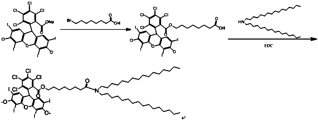
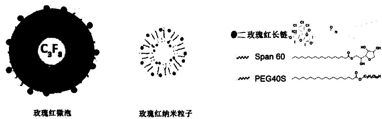
[0038] A preparation method of the microbubble preparation is as follows: figure 1 , shown in 2, includes the following steps:

[0039] (1) Tiger red sodium salt (0.5g, 4.92x 10 -4 mol), 8-bromooctanoic acid (0.33g, 1.48x 10 -3 mol) was added with 5mL DMF, stirred at 80°C for 7 hours, after the DMF in the product was spin-dried in vacuum, 50mL of ether was added, stirred at room temperature at 25°C for 18 hours, and filtered to obtain a red powder. Add 50mL of pure water to the red powder of the product, stir at room temperature 25 degrees Celsius for 18 hours, and filter to obtain product 1 0.44g, 3.95x10 -4 mol). The relative molecular mass and structure of the product were confirmed by mass spectrometry and NMR. 1 H NMR(500MHz,DMSO-d6):0.80(m,CH 2 ,2H),1.00(m,CH 2 ,2H),1.10(m,CH 2 ,4H),1.40(m,CH 2 ,2H),2.20(t,CH 2 COOH,2H),3.90(t,OCH 2 ,2H),7.50(s,ArH,2H),11.90(s,COOH,1H). Theoretical value of mass spectrum: 1115.8, experimental value 1114.6. The reaction formula...

Embodiment 2

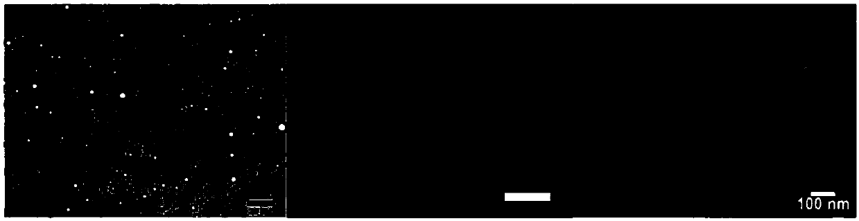
[0046] (1) The microbubble preparation prepared in Example 1 was observed under a fluorescence microscope to obtain imaging under white light and fluorescence images excited by 540nm green light.

[0047] (2) Then 1 mL of microbubbles was taken for ultrasonic crushing, dynamic light scattering and zeta potential evaluation were performed, and then the nanoparticles were observed under a transmission electron microscope to obtain image results.

[0048] The final result is as image 3 Shown, the fluorescence image of the microbubble preparation demonstrates the successful binding of rose bengal to the microbubbles, which become nanoparticles of the appropriate size after sonication.

Embodiment 3

[0050] The in vivo imaging effect evaluation of the microbubble preparation prepared in Example 1 mainly includes the following steps:

[0051] (1) Select a 2-2.5kg female New Zealand white rabbit, and remove the abdominal hair. Inject urethane via the marginal ear vein for anesthesia. Apply ultrasound coupling agent on the abdomen, and use ultrasound equipment to perform nephrography without contrast agent (frequency is 3.5MHz, mechanical index is 0.06 and 0.75). Save image.

[0052] (2) Inject 1 mL of microbubble preparation through the marginal ear vein, followed by injection of 4 mL of normal saline to better enter the blood circulation. They were then subjected to nephrography (frequency 5 MHz, mechanical index 0.06 and 0.75). Save image.

[0053] The result is as Figure 4 As shown, after the injection of rose bengal microbubbles, ultrasound contrast can be performed well under the condition of low mechanical index, which verifies its contrast function as microbubbl...


PUM


Abstract

The invention discloses a microbubble preparation for ultrasonic diagnosis and SDT (sonodynamic therapy) and a preparation method of the microbubble preparation. The microbubble preparation uses a rose-bengal long-chain compound and a surfactant as shell materials filled with gas, has the capabilities of ultrasonic imaging, ultrasonic located bursting and singlet oxygen production under ultrasonicstimulation, and can be used for tumor imaging and therapy. The drug loading capacity of microbubbles is improved effectively by drug synthesis and the microbubble preparation way, drug uptake of a tumor part is increased greatly by means of fixed-point bursting of the microbubbles by located ultrasound, and the tumor status is observed in real time through combination of ultrasonic imaging and therapy. The preparation can effectively increase diagnosis and treatment efficiency, is a high-efficiency novel drug loading system and has good clinical application prospects.

Description

technical field [0001] The invention belongs to the field of biomedical materials, and in particular relates to an ultrasonic contrast agent for ultrasonic imaging diagnosis and sonodynamic therapy and a preparation method thereof. Background technique [0002] Cancer has always been a problem that plagues the world, and its increasing morbidity and mortality are a major challenge to modern medicine. Chemotherapy is one of the important means of cancer treatment, but simple small molecule drugs will be rapidly metabolized by the liver and kidney after entering the body, resulting in a short half-life and residence time in the body, and the distribution of some drugs in the tumor is not high, in order to achieve therapeutic effect , may lead to drug concentrations in other organs reaching levels that are prone to toxicity. As an excellent drug delivery system, nanocarriers are gradually gaining attention. However, most nano-drug delivery systems utilize the EPR effect, alth...

Claims


Application Information

Patent Timeline
Patent Type & Authority Applications(China)
IPC IPC(8): A61K49/22A61K41/00A61K47/69A61P35/00
CPCA61K41/0033A61K47/6925A61K49/223A61P35/00
Inventor 王凡梁晓龙侯睿李小达史继云马晓途张旭
Owner PEKING UNIV
Why Eureka
Try Eureka