AI technical title is built by Patsnap AI team. It summarizes the technical point description of the patent document.
A technique for CT imaging and detection of objects
Pending Publication Date: 2017-07-07
TAISHAN MEDICAL UNIV
View PDF4 Cites 4 Cited by
Summary
Abstract
Description
Claims
Application Information
AI Technical Summary
This helps you quickly interpret patents by identifying the three key elements:
Problems solved by technology
Method used
Benefits of technology
Problems solved by technology
[0003] At present, the phantoms used for quality control of PET / CT imagers have problems such as single types and simple structures. The performance parameters of the CT part cannot perform quality control on the imaging quality of the combined use of PET / CT
Method used
Image
Click on the blue label to locate the original text in one second.
Reading with bidirectional positioning of images and text.
Examples
Embodiment 1
[0098] 1. The structure and material of the phantom body:
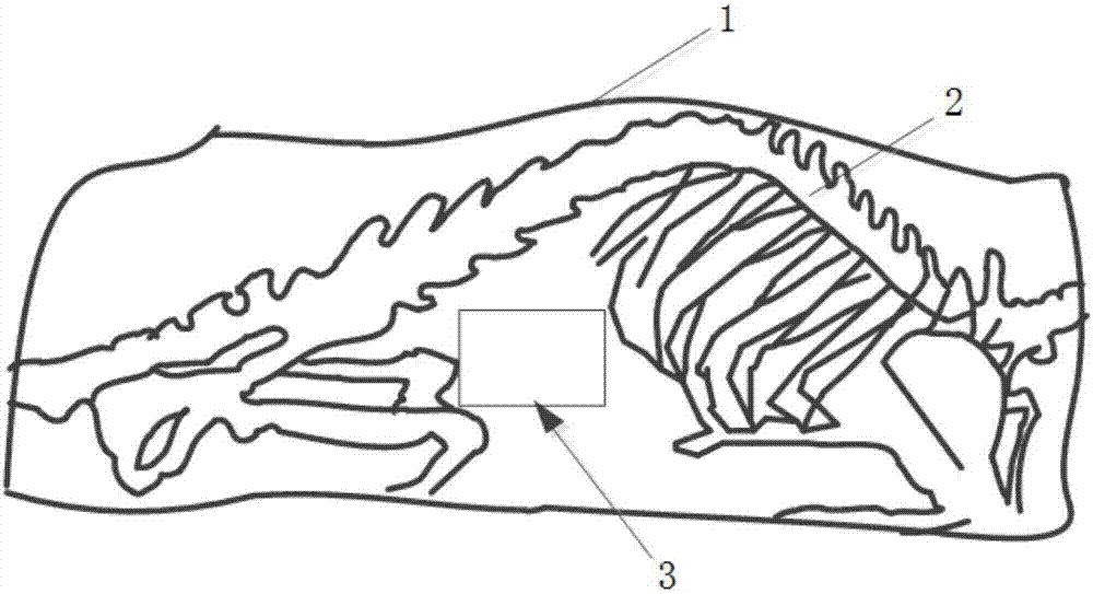
[0099] The overall shape of the phantom is that of a mouse. Specifically, a mouse model was constructed based on the CT scan data of real mice, and the mouse model was made by 3D printing technology according to the constructed model.
[0100] (1) Selection of model materials:
[0101] All skeletal structures of the mouse were 3D printed using bone tissue equivalent material 1; the skin and other internal tissues of the mouse were 3D printed using soft tissue equivalent material 2.
[0102] Among them, material 1 is a high-density resin with a Shorehardness greater than 90 among existing 3D printing materials.
[0103]Material 2 is EVA resin with lower density among existing 3D printing materials.
[0104] (2) The structure of the phantom:
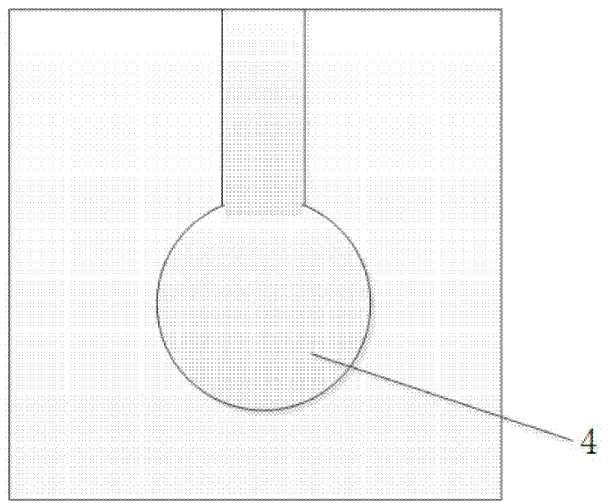
[0105] The phantom includes: a phantom body with a removable replacement module (for example, such as figure 1 As shown, located in the lower abdomen of the mouse model, avo...
Embodiment 2
[0109] 1. The structure and material of the phantom body:
[0110] The overall shape of the phantom is that of a mouse. Specifically, a mouse model was constructed based on the CT scan data of real mice, and the mouse model was made by 3D printing technology according to the constructed model.
[0111] (1) Selection of model materials:
[0112] All the bone structure of the mouse is printed with bone equivalent material 1, the skin of the mouse is made with material 3, and other tissues inside the mouse are infused with the prepared soft tissue equivalent material 4.
[0113] Material 1 is a high-density resin with a Shorehardness greater than 90 among existing 3D printing materials.
[0114] Material 3 is a common plastic material among existing 3D printing materials.
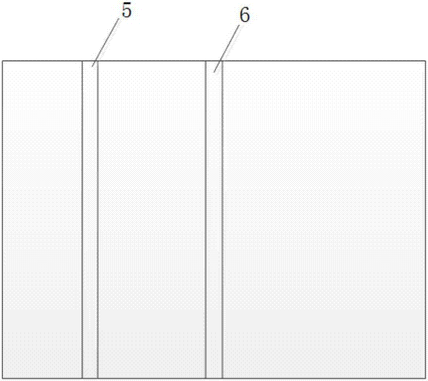
[0117] The phantom includes: a phantom body, and there are two detachable replacement modules in the phantom body (...
PUM
Abstract
The invention discloses an animal PET / CT (positron emission tomography / computed tomography) imaging quality detection phantom. The quality detection phantom comprises a phantom main body in the form of an animal, wherein at least one detachable substitute module is arranged in the phantom main body; and the detachable substitute module is selected from one or more of the following modules: an in-vivo tumor simulation module, a PET / CT image fusion degree test module, a PET / CT attenuation correction test module, a PET performance parameter test module and a CT performance parameter test module. The phantom provided by the invention is a quality detection phantom having morphological features; the phantom can simulate appearances, structures and focuses of real animals; the phantom is applicable to quality control of an animal PET / CT imaging instrument; and in addition, the phantom can be used for searching optimum scanning parameters of the PET / CT imaging instrument and is applicable to such aspects as teaching demonstration, tracer agent screening and the like; therefore, the phantom has a broad application scope.
Description
technical field [0001] The invention relates to the field of PET / CT quality assurance and quality control, in particular to an animal PET / CT imaging quality detection phantom. Background technique [0002] In recent years, with the rapid development of medical imaging technology, multimodal joint imaging technology has received extensive attention, such as PET / CT (Positron Emission Tomography / Computed Tomography, Positron Emission Tomography / X-rayComputed Tomography) Among them, PET can obtain functional and metabolic information very well, which belongs to functional imaging and has high sensitivity to diseased tissues, but its spatial resolution is low, and PET information cannot be accurately positioned; CT can provide high High-resolution images, but because they belong to anatomical imaging, they cannot reflect the functional changes of tissues well. The combination of PET and CT (PET / CT multimodal two-in-one imaging system) can make up for the shortcomings of PET ima...
Claims
Application Information
Patent Timeline
Application Date:The date an application was filed.
Publication Date:The date a patent or application was officially published.
First Publication Date:The earliest publication date of a patent with the same application number.
Issue Date:Publication date of the patent grant document.
PCT Entry Date:The Entry date of PCT National Phase.
Estimated Expiry Date:The statutory expiry date of a patent right according to the Patent Law, and it is the longest term of protection that the patent right can achieve without the termination of the patent right due to other reasons(Term extension factor has been taken into account ).
Invalid Date:Actual expiry date is based on effective date or publication date of legal transaction data of invalid patent.

