Blood smear self-focusing microimaging method

A technology of microscopic imaging and blood smear, which is applied in the field of self-focusing microscopic imaging of blood smear, and can solve problems such as focusing

Inactive Publication Date: 2017-07-28
SHENZHEN UNIV
View PDF6 Cites 14 Cited by
  • Summary
  • Abstract
  • Description
  • Claims
  • Application Information

AI Technical Summary

Problems solved by technology

[0005] The technical problem to be solved by the present invention is to provide a blood smear self-focusing microscopic

Method used


Image

  • Blood smear self-focusing microimaging method
  • Blood smear self-focusing microimaging method
  • Blood smear self-focusing microimaging method

Examples


Embodiment Construction

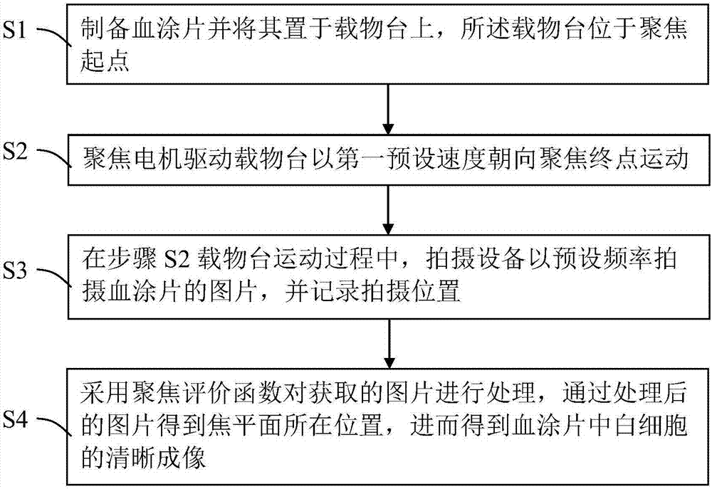
[0052] Such as figure 1 Shown is a schematic flow chart of an embodiment of the self-focusing microscopic imaging method for blood smears provided by the present invention, including a stage for placing blood smears, a focusing motor for driving the movement of the stage, and taking pictures of blood during the movement of the stage. The equipment for shooting smear pictures, the microscopic imaging method includes: S1 preparing a blood smear and placing it on the stage, the stage is located at the starting point of focus; S2 focusing motor drives the stage towards Focus end point movement; S3 During the movement of the stage in step S2, the shooting device takes pictures of the blood smear at a preset frequency and records the shooting position; S4 uses the focus evaluation function to process the acquired pictures, and through the processed pictures Get the position of the focal plane, and then get a clear image of the white blood cells in the blood smear.

[0053] In this ...


PUM


Abstract

The invention discloses a blood smear self-focusing microimaging method, which comprises an objective table for holding a blood smear, a focusing motor for driving the objective table to move, and a shooting device for shooting blood smear pictures in the moving process of the objective table. The method comprises the following steps: S1) the blood smear is prepared and placed on the objective table, wherein the objective table locates at a focusing start point; S2) the focusing motor drives the objective table to move to a focusing end point at a first preset speed; S3) in the moving process of the objective table in the step S2), the shooting device shoots the blood smear pictures with a preset frequency and records the shooting position; and S4) the obtained pictures are subjected to processing through a focusing evaluation function, and the position where a focal plane locates is obtained through the processed pictures to obtain clear imaging of leucocytes in the blood smear. The pictures shot by the shooting device are processed through the focusing evaluation function, and imaging of the blood smear is obtained according to the processed images, so that focusing can be realized without labor, and manpower and material resources are saved.

Description

technical field [0001] The invention relates to the field of imaging technology, in particular to a blood smear self-focusing microscopic imaging method. Background technique [0002] Blood smear examination is an important method for diagnosing many diseases. Information on the number, shape, and proportion of white blood cells in blood smears can help doctors diagnose diseases. Therefore, accurate and rapid classification and counting of white blood cells is a direct technical problem faced by the medical field today, and it is also the primary problem for hospitals to improve work efficiency. [0003] At present, hospitals generally test the type and quantity of white blood cells in the blood through blood analyzer method and microscope method. The blood analyzer method has the advantages of fast speed, good repeatability, and high accuracy, but it cannot accurately identify cell morphology and its pathological changes. Once abnormal patients are found, further examinati...

Claims


Application Information

Patent Timeline
IPC IPC(8): G02B21/00G02B21/34G02B21/36
CPCG02B21/006G02B21/34G02B21/361G02B21/367
Inventor 李乔亮钟金亮齐素文陈哲玮谢林培张倩许育祯吴亚杰
Owner SHENZHEN UNIV
Why Eureka
Try Eureka