Method and device for extracting pulmonary blood vessels from pectoral CT (computed tomography) images

A CT image, blood vessel technology, applied in the computer field

Active Publication Date: 2017-08-15
NORTHEASTERN UNIV
View PDF5 Cites 13 Cited by
  • Summary
  • Abstract
  • Description
  • Claims
  • Application Information

AI Technical Summary

Problems solved by technology

Moreover, clinicians can only check the lesion area through layer-by-layer image observation

Method used


Image

  • Method and device for extracting pulmonary blood vessels from pectoral CT (computed tomography) images
  • Method and device for extracting pulmonary blood vessels from pectoral CT (computed tomography) images
  • Method and device for extracting pulmonary blood vessels from pectoral CT (computed tomography) images

Examples


Embodiment Construction

[0072] The technical solutions in the embodiments of the present invention will be clearly and completely described below in conjunction with the accompanying drawings in the embodiments of the present invention. Obviously, the described embodiments are only some, not all, embodiments of the present invention. Based on the embodiments of the present invention, all other embodiments obtained by persons of ordinary skill in the art without making creative efforts belong to the protection scope of the present invention.

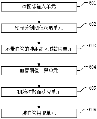
[0073] The present invention considers that the operation is very difficult if the pulmonary vessels are directly extracted from the chest CT images due to the small differences in the gray values ​​of the heart, surrounding bones and pulmonary vessels in the chest CT images. Therefore, the present invention first uses the method of preset segmentation threshold and 3D area growth to obtain lung tissue without blood vessels, and then obtains the closed lung tissu...


PUM


Abstract

The present invention discloses a method and device for extracting pulmonary blood vessels from pectoral CT (computed tomography) images. The method includes the following steps that: n layers of inputted pectoral CT images are received, a designated image layer is obtained and is subjected to gray-scale mapping, so that a preset segmentation threshold value can be obtained; designated pixel points of a lung region are selected from the designated image layer, so that initial seed points can be obtained, 3D regional growth is performed according to the preset segmentation threshold value and the initial seed points, and a lung tissue region without blood vessels is obtained; morphological operation is performed on the lung tissue region, so that a closed lung tissue region with blood vessels is obtained, and a blood vessel threshold value is calculated; pixel points larger than the blood vessel threshold value are searched in the closed lung tissue region with blood vessels, and the pixel points are adopted as initial mark points, and an initial diffusion surface is obtained; and surface diffusion is performed in the closed lung tissue region with blood vessels according to the initial diffusion surface and the blood vessel threshold value, so that pulmonary blood vessels can be obtained. With the method and device of the invention adopted, the pulmonary blood vessels can be accurately extracted from the pectoral CT images, and therefore, doctors can be assisted to diagnose pulmonary vascular disease accurately.

Description

technical field [0001] The invention relates to the field of computer technology, in particular to a method and device for extracting pulmonary blood vessels from chest CT images. Background technique [0002] CT is the abbreviation of computer X-ray tomography technology, which is a commonly used medical imaging equipment. CT images are black and white images, with different gray levels representing the degree of absorption of X-rays by organs and tissues. For example, on a chest CT image, areas of low density (that is, low gray value) represent trachea and lung parenchyma, and areas of high density (that is, high gray value) represent blood vessels, chest cavity, bones, etc. CT can intuitively display the lesion area on the image, which provides a convenient and reliable basis for doctors to observe and diagnose diseases. Usually, CT images are cross-sectional images, and in order to display the entire organ, multiple consecutive slice images are required. [0003] In t...

Claims


Application Information

Patent Timeline
Patent Type & Authority Applications(China)
IPC IPC(8): G06T7/11G06T7/136
CPCG06T7/0012G06T2207/10081G06T2207/30061
Inventor 覃文军路石洁赵姝颖杨金柱赵大哲栗伟曹鹏冯朝路宫照煊边子健姚洪柱陈世伟孙强
Owner NORTHEASTERN UNIV
Why Eureka
Try Eureka