Scanning method and device for positron emission tomography (PET) equipment

A computer equipment and positron emission technology, applied in the field of image processing, can solve the problems of affecting the quality of image reconstruction, inaccurate phase segmentation, unable to remove abnormal data, etc., and achieve the effect of improving the quality of reconstructed images and the method is simple

Active Publication Date: 2017-08-18
SHENYANG INTELLIGENT NEUCLEAR MEDICAL TECH CO LTD
View PDF6 Cites 8 Cited by
  • Summary
  • Abstract
  • Description
  • Claims
  • Application Information

AI Technical Summary

Problems solved by technology

[0003] During the process of implementing this application, the applicant found that with the attenuation of the tracer and the occurrence of equipment abnormalities, some data that may affect the quality of image reconstruction may be generated during the scanning process
In addition, in cardiac and respiratory gated scans, abnormal heart or respiratory fluctuations may cause inaccurate phase segmentation and affect the quality of reconstructed images
In the prior art, before image reconstruction, the original data is compressed, and the original data is compressed and divided into several data segments. Even if an abnormality occurs in the image reconstruction, the original data cannot be restored, so the abnormal data cannot be eliminated. Have the patient rescanned and use the data from the rescan for a second image reconstruction
Therefore, the scanning method provided by the prior art has the defects of cumbersome operation, inflexibility, and inability to remove abnormal data

Method used


Image

  • Scanning method and device for positron emission tomography (PET) equipment
  • Scanning method and device for positron emission tomography (PET) equipment
  • Scanning method and device for positron emission tomography (PET) equipment

Examples


Embodiment Construction

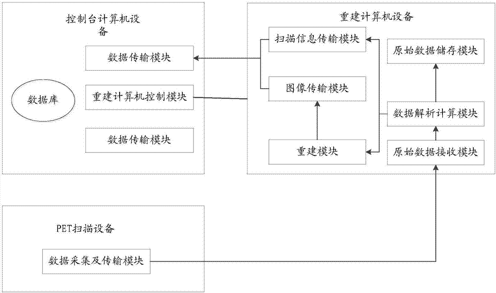
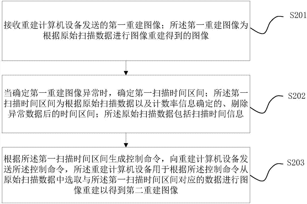
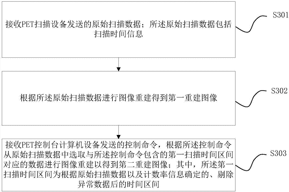
[0023] The embodiment of the present application provides a scanning method and device for PET equipment. When it is determined that the first reconstructed image is abnormal, the scanning time interval in which the abnormal data is removed can be re-determined, and the image reconstruction is performed according to the data corresponding to the scanning time interval to obtain The second reconstructed image effectively improves the quality of the reconstructed image, and the method is simple, flexible and effective.

[0024] Terms used in the embodiments of the present application are only for the purpose of describing specific embodiments, and are not intended to limit the present application. The singular forms "a", "said" and "the" used in the embodiments of this application and the appended claims are also intended to include plural forms unless the context clearly indicates otherwise. It should also be understood that the term "and / or" as used herein refers to and includ...


PUM


Abstract

An embodiment of the invention provides a scanning method for PET equipment, which is applied to PET console computer device. The scanning method comprises the following steps: receiving a first reconstructed image transmitted by reconstruction computer equipment, wherein the first reconstructed image is an image obtained by performing image reconstruction based on original scanning data; determining a first scanning time interval when determining that the first reconstructed image is abnormal, wherein the first scanning time interval is a time interval determined based on the original scanning data and counting rate information after abnormal data are deleted, and the original scanning data comprise scanning time information; generating a control command according to the first scanning time interval; and transmitting the control command to the reconstruction computer equipment, wherein the reconstruction computer equipment is used for selecting data corresponding to the first scanning time interval from the original scanning data according to the control command to reconstruct an image and obtain a second reconstructed image. The embodiment of the invention can effectively improve the quality of the reconstructed image; the method is simple, flexible and effective.

Description

technical field [0001] The embodiments of the present application relate to the technical field of image processing, and in particular to a scanning method and device for a PET device for positron emission computed tomography. Background technique [0002] At present, Positron Emission Tomography (abbreviated as PET in English) equipment has been widely used in the medical field. PET is a medical imaging technology that uses positron nuclide as a tracer, reflects its metabolic changes through the uptake of the tracer by human body parts, and obtains in-vivo images by scanning with a positron electron scanner. Generally speaking, the metabolism of the tumor lesion is abnormal, and the uptake of the tracer is also abnormal, so the state of the lesion can be judged accordingly. [0003] During the process of implementing the present application, the applicant found that with the attenuation of the tracer and the occurrence of equipment abnormalities, some data that may affect ...

Claims


Application Information

Patent Timeline
Patent Type & Authority Applications(China)
IPC IPC(8): A61B6/03
CPCA61B6/037A61B6/486A61B6/5211
Inventor 孙奇李明吕斌
Owner SHENYANG INTELLIGENT NEUCLEAR MEDICAL TECH CO LTD
Why Eureka
Try Eureka