Virtual endoscopy system based on CT image

A virtual endoscope and CT image technology, applied in the field of medical image processing systems, can solve problems such as the overall composition and operation of the virtual endoscope system that have not been revealed, and achieve the goal of improving visual fidelity, increasing authenticity, and reducing inspection difficulty Effect

Pending Publication Date: 2017-09-26
THE AFFILIATED HOSPITAL OF QINGDAO UNIV
View PDF6 Cites 7 Cited by
  • Summary
  • Abstract
  • Description
  • Claims
  • Application Information

AI Technical Summary

Problems solved by technology

However, the data processing method disclosed in this patent application does not reveal the overall composition and operation of the virtual endoscopy system

Method used


Image

  • Virtual endoscopy system based on CT image

Examples


Embodiment Construction

[0025] Embodiments of the present invention are described in detail below, examples of which are shown in the drawings, wherein the same or similar reference numerals designate the same or similar elements or elements having the same or similar functions throughout. The embodiments described below by referring to the figures are exemplary and are intended to explain the present invention and should not be construed as limiting the present invention.

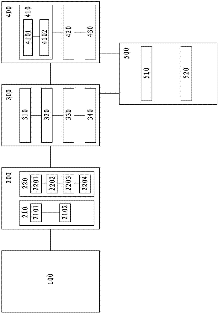
[0026] Please refer to figure 1, as a non-limiting implementation, the CT image-based virtual endoscopy system of the present invention includes: an image acquisition module 100, an image processing module 200, a three-dimensional transformation module 300, a centerline extraction module 400 and a roaming module 500.

[0027] The image acquisition module 100 receives CT images in DICOM format taken from different sections from a specific organ.

[0028] The image processing module 200 includes an image preprocessing unit 210 and...


PUM


Abstract

The present invention discloses a virtual endoscopy system based on a CT image. The system comprises: an image collection module configured to receive a plurality of CT images; an image processing module being in communication connection with the image collection module and configured to perform smooth processing and boundary processing of each CT image in the CT images transmitted by the image collection module; a three-dimensional conversion module being in communication connection with the image processing module and configured to perform surface reconstruction of each CT image after the processing through the image processing module to obtain a special three-dimensional image; a center line extraction module being in communication connection with the three-dimensional conversion module and configured to extract the center line of the special three-dimensional image; and a roaming module being in communication connection with the three-dimensional conversion module and the center line extraction module and configured to control the movement track of a virtual camera taking a center line as a viewpoint in the special three-dimensional image and perform roaming.

Description

technical field [0001] The invention relates to a medical image processing system, in particular to a medical image three-dimensional visualization system. Background technique [0002] With the development of modern medical imaging technology, modern medical equipment can help doctors diagnose internal organ or tissue lesions of patients without surgery. Computed tomography (CT) and magnetic resonance imaging (magnetic resonance MR) can produce data representing information about the internal organs of the human body. However, these data are often not given in a continuous three-dimensional form, they only form a series of tissue or organ sections, so only experienced doctors can correctly reconstruct the patient's internal organs and tissues, which gives Diagnosis introduces a great deal of subjectivity. [0003] Virtual endoscopy (Virtual endoscopy, VE) technology is a unique technology gradually formed with the intersection and integration of medical imaging technology...

Claims


Application Information

Patent Timeline
Patent Type & Authority Applications(China)
IPC IPC(8): G06T19/00G06T7/00A61B6/03A61B6/00
CPCA61B6/032A61B6/466A61B6/5211G06T7/0012G06T19/003G06T2207/10081
Inventor 魏宾董蒨卢云朱呈瞻董冰子于綦悦夏楠
Owner THE AFFILIATED HOSPITAL OF QINGDAO UNIV
Why Eureka
Try Eureka