Method of identifying edema and hematoma in MRI image and apparatus thereof

An image-in-image technique used to identify edema and hematoma in MRI images

Active Publication Date: 2017-10-24
BEIJING SHENRUI BOLIAN TECH CO LTD +1
View PDF10 Cites 20 Cited by
  • Summary
  • Abstract
  • Description
  • Claims
  • Application Information

AI Technical Summary

Problems solved by technology

Brain lesions include cerebral edema and cerebral hematoma, but in general, hematoma will be accompanied by edema, and the hematoma mass is often wrapped in the edema mass. In this case, it is difficult for doctors to distinguish based on experience. Edema and hematoma

Method used


Image

  • Method of identifying edema and hematoma in MRI image and apparatus thereof
  • Method of identifying edema and hematoma in MRI image and apparatus thereof
  • Method of identifying edema and hematoma in MRI image and apparatus thereof

Examples


Embodiment Construction

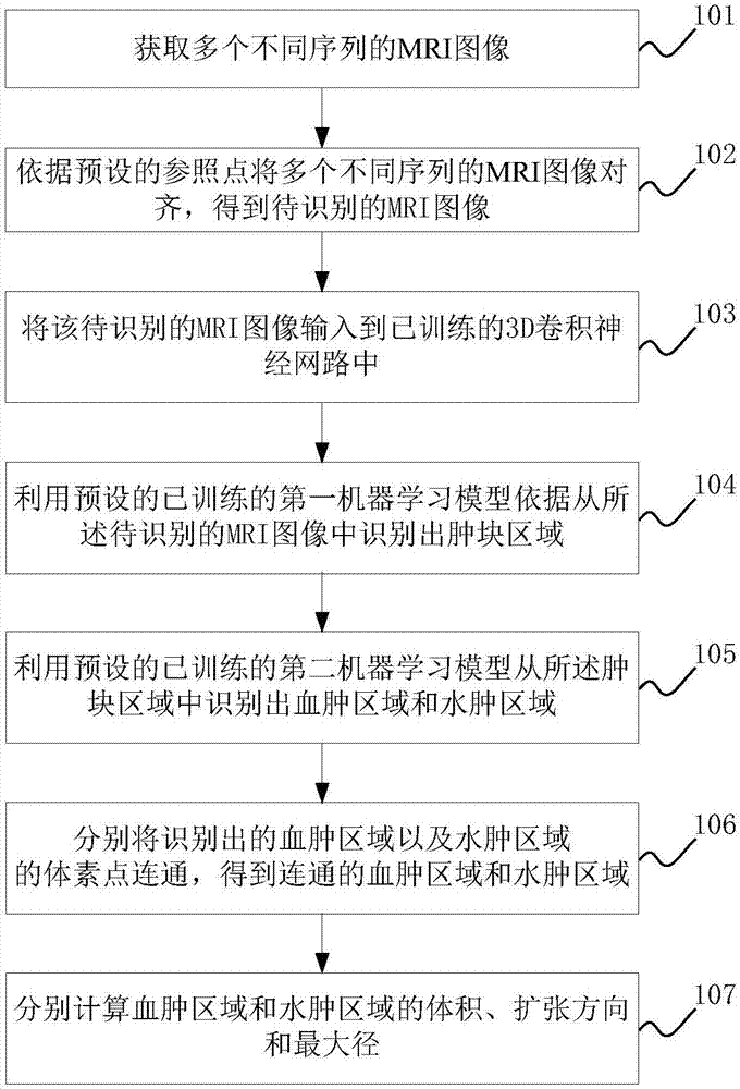
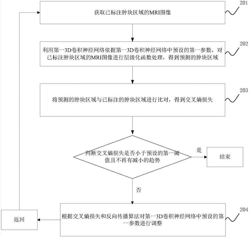
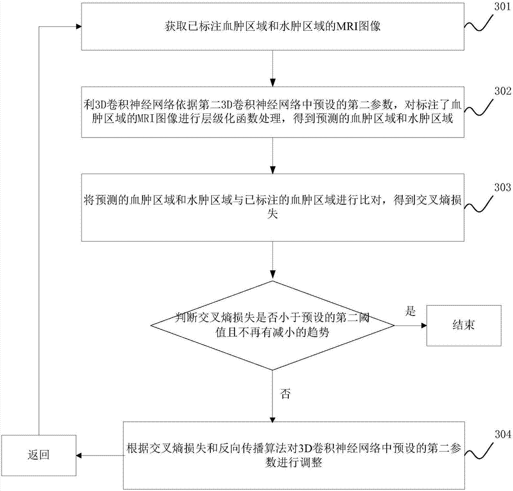
[0064] The method for identifying edema and hematoma proposed in the embodiments of the present application is applied to MRI images, with the purpose of automatically identifying edema and hematoma from MRI images.

[0065] The method for identifying edema and hematoma described in the embodiment of the present application may be performed by a device for identifying edema and hematoma, and the device may be integrated into existing MRI equipment or set independently. In the case of a stand-alone setup, MRI images can be acquired from existing MRI scanning equipment.

[0066] The following will clearly and completely describe the technical solutions in the embodiments of the present invention with reference to the accompanying drawings in the embodiments of the present invention. Obviously, the described embodiments are only some, not all, embodiments of the present invention. Based on the embodiments of the present invention, all other embodiments obtained by persons of ordi...


PUM


Abstract

The invention discloses a method of identifying an edema and a hematoma in a MRI image and an apparatus thereof. The method is characterized by firstly, using a first preset machine learning model to identify a lump area in the MRI image to be identified, wherein the first preset machine learning model is acquired after training the MRI image where the lump area is marked; then, using a second preset machine learning model to identify a hematoma area and an edema area in the lump area, wherein the second preset machine learning model is acquired after training the MRI image where the hematoma area and the edema area are marked; and calculating sizes, expansion directions and maximum diameters of the hematoma area and the edema area. By using the method, the hematoma and the edema can be automatically and high-efficiently identified; and the sizes, the expansion directions and the maximum diameters of the hematoma area and the edema area can be automatically calculated so that a doctor is helped to accurately determine a condition.

Description

technical field [0001] The invention relates to the field of medical image processing, in particular to a method and device for identifying edema and hematoma in MRI images. Background technique [0002] With the development of digital medical technology, the role of medical images in the medical diagnosis process has become more and more obvious, and it has gradually become the main tool for doctors to observe human lesions. When diagnosing a disease, doctors generally search for regions of interest representing lesions from medical images artificially based on experience. [0003] MRI (English full name: Magnetic Resonance Imaging, Chinese full name: Nuclear Magnetic Resonance Imaging) image is one of the commonly used medical images. MRI imaging is a kind of tomographic imaging. It uses magnetic resonance to obtain electromagnetic signals from the human body and reconstruct the human body. information to obtain MRI images. The application of MRI images in the nervous sy...

Claims


Application Information

Patent Timeline
Patent Type & Authority Applications(China)
IPC IPC(8): G06T7/00G06T7/187G06T7/38
CPCG06T7/0012G06T7/187G06T7/38G06T2207/10088G06T2207/20081G06T2207/20084G06T2207/30016
Inventor 张番栋周振庞亮张树孔明王洪振李一鸣
Owner BEIJING SHENRUI BOLIAN TECH CO LTD
Why Eureka
Try Eureka