
DR (direct digital radiograph) dual-energy subtraction high-KV (kilovolt) chest radiography method for pneumoconiosis diagnosis

A dual-energy subtraction and pneumoconiosis technology, which is applied in the fields of radiodiagnostic equipment, diagnosis, and clinical application of radiodiagnosis, can solve the problems of DR dual-energy subtraction high-KV radiography screening and diagnosis that have not yet been seen. , to achieve the effect of rich image display layers, large exposure latitude, and clear display

Inactive Publication Date: 2017-11-03
合肥市第三人民医院
View PDF2 Cites 1 Cited by
  • Summary
  • Abstract
  • Description
  • Claims
  • Application Information

AI Technical Summary

Problems solved by technology

[0005] At present, there are no corresponding reports and literatures on the use of DR dual-energy subtraction high-KV radiography for the screening and diagnosis of pneumoconiosis

Method used


Image

  • DR (direct digital radiograph) dual-energy subtraction high-KV (kilovolt) chest radiography method for pneumoconiosis diagnosis
  • DR (direct digital radiograph) dual-energy subtraction high-KV (kilovolt) chest radiography method for pneumoconiosis diagnosis
  • DR (direct digital radiograph) dual-energy subtraction high-KV (kilovolt) chest radiography method for pneumoconiosis diagnosis

Examples


Embodiment Construction

[0019] The DR dual energy subtraction high KV chest radiography method for the diagnosis of pneumoconiosis includes the following steps:

[0020] (1) Posterior-anterior chest radiographs for those with a history of dust exposure;

[0021] (2) The DR system uses the double exposure method:

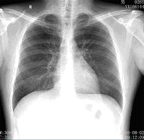
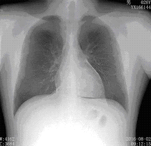
[0022] The exposure interval time is 100ms, and the output energy of the X-ray tube for the two exposures is 70 kV and 120 kV respectively. figure 1 common DR image, such as figure 2 The three images of pure lung tissue image and pure bone tissue image; set the position of the tube, its center line is aligned with the level of the sixth thoracic vertebra of the screening object, and the human body maintains the same position of the conventional chest posteroanterior position, and maintains the position after fully inhaling Breath-holding state.

[0023] (3) According to the above three images, the diagnosis of pneumoconiosis was carried out according to the diagnostic criteria of GBZ 70...


PUM


Abstract

The invention discloses a DR (direct digital radiograph) dual-energy subtraction high-KV (kilovolt) chest radiography method for pneumoconiosis diagnosis. Compared with a traditional high-KV radiography method, the DR dual-energy subtraction high-KV chest radiography method has the advantages that first-grate radiography rate and small shadow detection rate in the lung can be remarkably increased when applied to pneumoconiosis screening and diagnosis, small dust shadows and subtle structural shadows thereof can be seen clearly through 120-KV exposure, and important clinical significance is achieved to pneumoconiosis diagnosis especially for early diagnosis.

Description

technical field [0001] The present invention relates to the application of digital X-ray photography (Direct Digital Radiography, DR) technology, in particular to the application of DR dual-energy subtraction chest radiography method in the diagnosis of pneumoconiosis. Background technique [0002] Pneumoconiosis is the accumulation of a large amount of dust in the lung tissue, causing pathological changes of lung tissue fibrosis and progressive damage to the lung tissue. Patients with pneumoconiosis have no specific symptoms in the early stage. As the disease progresses, most of them will develop cough, sputum, hemoptysis, chest pain, dyspnea, systemic symptoms and even serious complications. Pneumoconiosis is the most important occupational disease in my country. It not only has a large number of patients, but also has great harm. It is a disease that reduces labor capacity, disables people, and affects life expectancy. It is also a major occupational disease compensated b...

Claims


Application Information

Patent Timeline
Patent Type & Authority Applications(China)
IPC IPC(8): A61B6/00
CPCA61B6/00A61B6/50A61B6/542
Inventor 胡茂能
Owner 合肥市第三人民医院
Learn More