Computer aided method for histological grading of breast invasive ductal carcinoma

A computer-aided, histological technology, applied in computing, image analysis, image data processing, etc., can solve the problems of consumption of computing resources, low error rate of detection and recognition, failure to meet accuracy requirements, etc., and achieve the effect of saving computing resources

Inactive Publication Date: 2017-11-07
成都知识视觉科技有限公司 +1
View PDF6 Cites 22 Cited by
  • Summary
  • Abstract
  • Description
  • Claims
  • Application Information

AI Technical Summary

Problems solved by technology

[0008] 2. Cellular atypia
[0014] However, the current mainstream histological grading method for invasive ductal carcinoma of the breast is based on the Nottingham scoring system, and the three indicators of the degree of duct formation, nuclear pleomorphism and mitotic count are respectively image labeled, classification model training and Detection and recognition, although this method imitates the doctor's diagnostic method, but because the detection and recognition of each indicator has a high error rate, it is currently unable to meet the accuracy requirements of clinical detection
At the same time, this method will consume a lot of computing resources

Method used


Image

  • Computer aided method for histological grading of breast invasive ductal carcinoma
  • Computer aided method for histological grading of breast invasive ductal carcinoma

Examples


Embodiment 1

[0033] Embodiment 1: a kind of computer-aided method for the histological grading of breast invasive ductal carcinoma, comprises the steps:

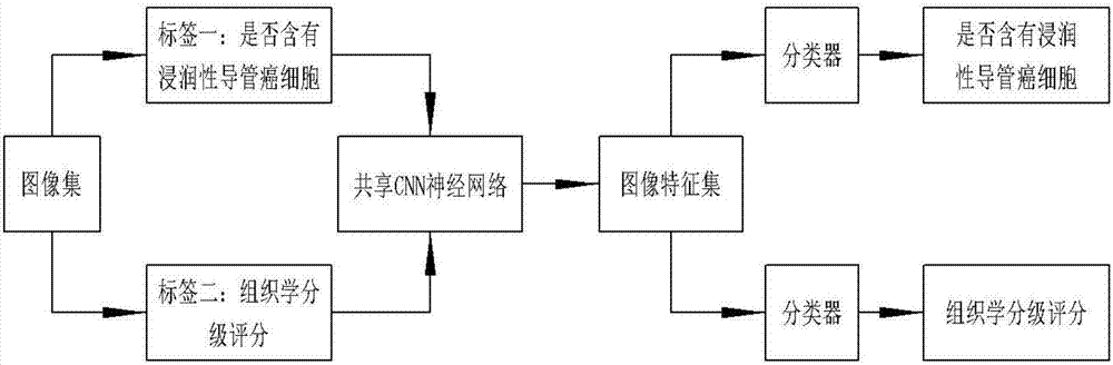
[0034] A. The pathologist manually marks the area where breast invasive ductal carcinoma cells exist in the digital slice image of breast cancer histology;

[0035] B. According to the Nottingham histological grading system, the pathologist gives a histological grading score to the area marked as invasive ductal carcinoma of the breast;

[0036] C. The computer reads in the digital slice image file that has been marked and histologically graded, cuts the image into small pieces of images, and obtains the label information of each small piece of image by querying the information of invasive ductal carcinoma region labeling and histological grading and scoring , the label information includes whether it contains invasive ductal carcinoma cells and histological grading score, and two types of sample sets are obtained, namely: a. sample sets...

Embodiment 2

[0039] Embodiment 2: a kind of computer-aided method for the histological grading of breast invasive ductal carcinoma, comprises the steps:

[0040] A. The pathologist manually marks the area where breast invasive ductal carcinoma cells exist in the digital slice image of breast cancer histology;

[0041] B. According to the Nottingham histological grading system, the pathologist gives a histological grading score to the area marked as invasive ductal carcinoma of the breast;

[0042]C. The computer reads in the digital slice image file that has been marked and histologically graded, cuts the image into fixed-size small block images, and obtains the information of each small block image by querying the information of invasive ductal carcinoma region labeling and histological grading. Label information to obtain two types of sample sets, namely: a. sample sets containing invasive ductal carcinoma cells; b. sample sets not containing invasive ductal carcinoma cells;

[0043] D....

Embodiment 3

[0045] Embodiment 3: a kind of computer-aided method for the histological grading of breast invasive ductal carcinoma, comprises the steps:

[0046] 1) The pathologist selects the area containing invasive ductal carcinoma in the breast cancer digital slice (Whole Slide Image, WSI) for manual labeling and assigns a histological grade according to the Nottingham histological grading system;

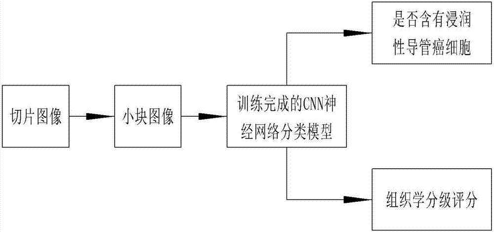
[0047] 2) Cut the image into small patches (patches), such as: the size of each patch is 256x256 pixels, and obtain whether the patch contains invasive ductal carcinoma and histological grade information by querying the information in the pathologist's annotation file , so as to obtain the label information of each small block image, and obtain two types of sample sets, namely: a. sample sets containing invasive ductal carcinoma cells, b. sample sets without invasive ductal carcinoma cells;

[0048] 3) In the sample set containing invasive ductal carcinoma cells, the histological grading la...


PUM


Abstract

The invention discloses a computer aided method for histological grading of the breast invasive ductal carcinoma. Pathologists manually label the region where breast invasive ductal carcinoma cells exist in a breast carcinoma histological digital slice image, and a histological grading score of the region which is labeled as a breast invasive ductal carcinoma region is given; the digital slice image file of which labeling and histological grading are completed is read in, the image is cut into small image blocks, by querying invasive ductal carcinoma region labels and histological grading score information, tag information of each small image block is obtained, and two kinds of sample sets are obtained; the CNN nerve network is made use of, multiple tags are built, classification based training tasks are learned deeply, the nerve network is allowed to automatically learn image features of the small image blocks, and a classification model is obtained; the classification model is made use of, the small image blocks extracted out from the breast carcinoma histological slice image is classified and identified.

Description

technical field [0001] The invention relates to a computer-aided method for histological grading of breast invasive ductal carcinoma. Background technique [0002] The female mammary gland is composed of skin, fibrous tissue, mammary glands and fat. Breast cancer is a malignant tumor that occurs in the mammary gland epithelial tissue. 99% of breast cancer occurs in women, and only 1% in men. Breast cancer is the number one common malignant tumor in women. [0003] Breast cancer is the most common cancer among women in China, as in most other countries. Breast invasive ductal carcinoma (IDC), non-specific type (NST) is the most common type of breast cancer. It originates from the mammary duct, and the cancer cells break through the duct wall and infiltrate into the breast interstitium and adipose tissue. Therefore, IDC can pass through Spread (metastasis) to other parts of the body through the lymphatic system or blood. IDC accounts for the vast majority of breast cancer, ...

Claims


Application Information

Patent Timeline
Patent Type & Authority Applications(China)
IPC IPC(8): G06F19/00G06T7/00
CPCG06T7/0012G06T2207/20084G06T2207/30068G06T2207/30096
Inventor 包骥向飞
Owner 成都知识视觉科技有限公司
Why Eureka
Try Eureka