

Micro-fluidic chip and research method for capturing and detecting exosome
A microfluidic chip and exosome technology, applied in chemical instruments and methods, measuring devices, laboratory containers, etc., can solve the problems that capture and detection cannot be carried out at the same time and consume a large amount of samples.
- Summary
- Abstract
- Description
- Claims
- Application Information
AI Technical Summary
Problems solved by technology
Method used
Image

Examples
Embodiment 1
[0032] The microfluidic chip used was designed and manufactured by our laboratory.
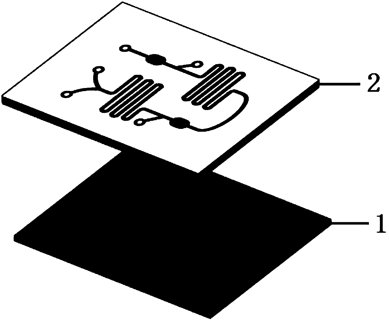
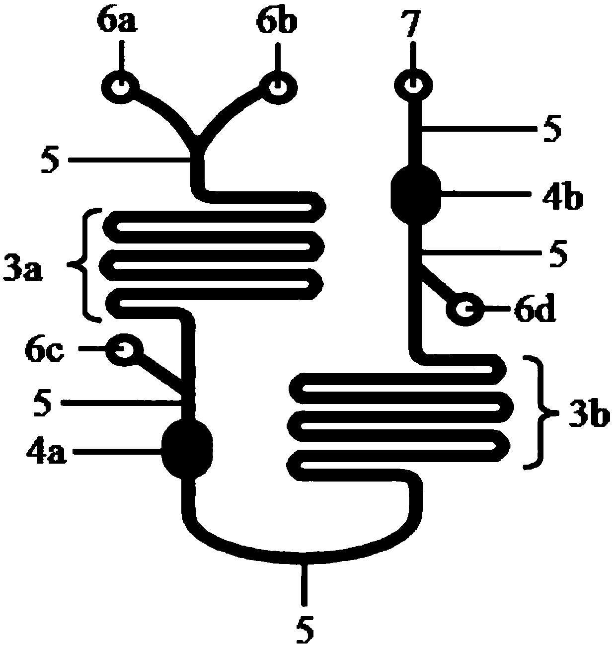
[0033] Such as Figure 1-3 As shown, the microfluidic chip provided by the present invention is composed of a glass substrate 1 and a membrane 2, the material of the membrane 2 is polydimethylsiloxane (PDMS), and the PDMS membrane 2 and the The glass substrate 1 is sealed. The PDMS membrane 2 is provided with a curved mixing channel, a collection pool, a connecting channel, a sample inlet and a liquid outlet;
[0034] Wherein, the first sample inlet 6a and the second sample inlet 6b are connected with the first curved mixing channel 3a through the connecting channel 5; the first curved mixing channel 3a is connected with the first collection pool 4a through the connecting channel 5, and in A third sample inlet 6c is provided between the first curved mixing channel 3a and the first collection tank 4a, and the third sample inlet 6c is opened on the side of the connecting channel 5; The curved...
Embodiment 2
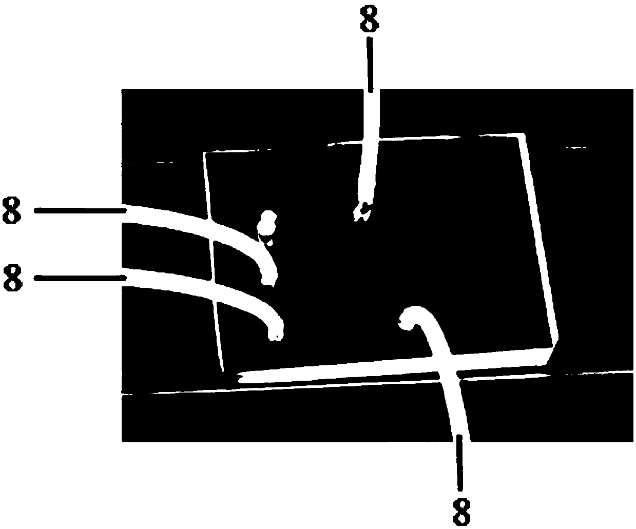
[0037] The chip described in Example 1 is used for research. The operation process of the chip is as follows: firstly, the magnetic nanoparticles with the anti-exosome surface antigen antibody attached to the surface are added to the first injection port 6a, and the blank cell culture solution is added to the first injection port 6a. Two sample inlets 6b, the small magnet 9 is placed on the first collecting pool 4a place ( Figure 4); In the second step, two syringe pumps 8 are respectively connected with the first injection port 6a and the second injection port 6b, and the magnetic nanoparticles and the culture solution are driven to flow through the first injection port of the chip at a flow rate of 2 microliters / minute. A curved mixing channel 3a; the third step is to investigate under a microscope whether the nanoparticles can be enriched in the collection pool 4a (for details, see Figure 5 ).
Embodiment 3
[0039] Adopt the chip described in embodiment 1 to carry out research, the operating process of described chip is:
[0040] First, magnetic nanoparticles linked with CD63 antibody were used to shake the culture medium of normal fibroblasts, MCF7, MDA-MB-231, SACC-83, and SACC-LM cells overnight at 4°C to make exosomes Fully combined with the CD63 antibody on the surface of the immunomagnetic nanoparticles, and the culture medium without cultured cells was used as the control;
[0041] In the second step, the mixture of immunomagnetic nanoparticles and exosomes is added to the first injection port 6a, and the detection primary antibody MHC I is added to the second injection port 6b, and the syringe pump 8 is used to drive the liquid to flow in the channel of the chip. The first curved mixing pool 3a is fully mixed to form a complex A of immunomagnetic nanoparticles, exosomes and MHC I;
[0042] In the third step, a small magnet 9 is placed under the first collection pool 4a to...
PUM
| Property | Measurement | Unit |
|---|---|---|
| Length | aaaaa | aaaaa |
| Width | aaaaa | aaaaa |
| Depth | aaaaa | aaaaa |
Abstract
Description
Claims
Application Information