Image display method, device and system for transanal endoscopic microsurgery navigation

An image display device and a technology for minimally invasive surgery, applied in the medical field, can solve problems such as limited auxiliary information, achieve the effects of occlusion and clear judgment before and after, reduce computational complexity, and smooth transition

Inactive Publication Date: 2018-01-19
ARIEMEDI MEDICAL SCI BEIJING CO LTD
View PDF4 Cites 9 Cited by
  • Summary
  • Abstract
  • Description
  • Claims
  • Application Information

AI Technical Summary

Problems solved by technology

However, the auxiliary information provided to doctors in the three views of

Method used


Image

  • Image display method, device and system for transanal endoscopic microsurgery navigation
  • Image display method, device and system for transanal endoscopic microsurgery navigation
  • Image display method, device and system for transanal endoscopic microsurgery navigation

Examples


Embodiment Construction

[0047] Embodiments of the present invention are described below with reference to the drawings. Elements and features described in one drawing or one embodiment of the present invention may be combined with elements and features shown in one or more other drawings or embodiments. It should be noted that representation and description of components or processes that are not relevant to the present invention and known to those of ordinary skill in the art are omitted from the drawings and descriptions for the purpose of clarity.

[0048] The present invention will be further described below in conjunction with the accompanying drawings.

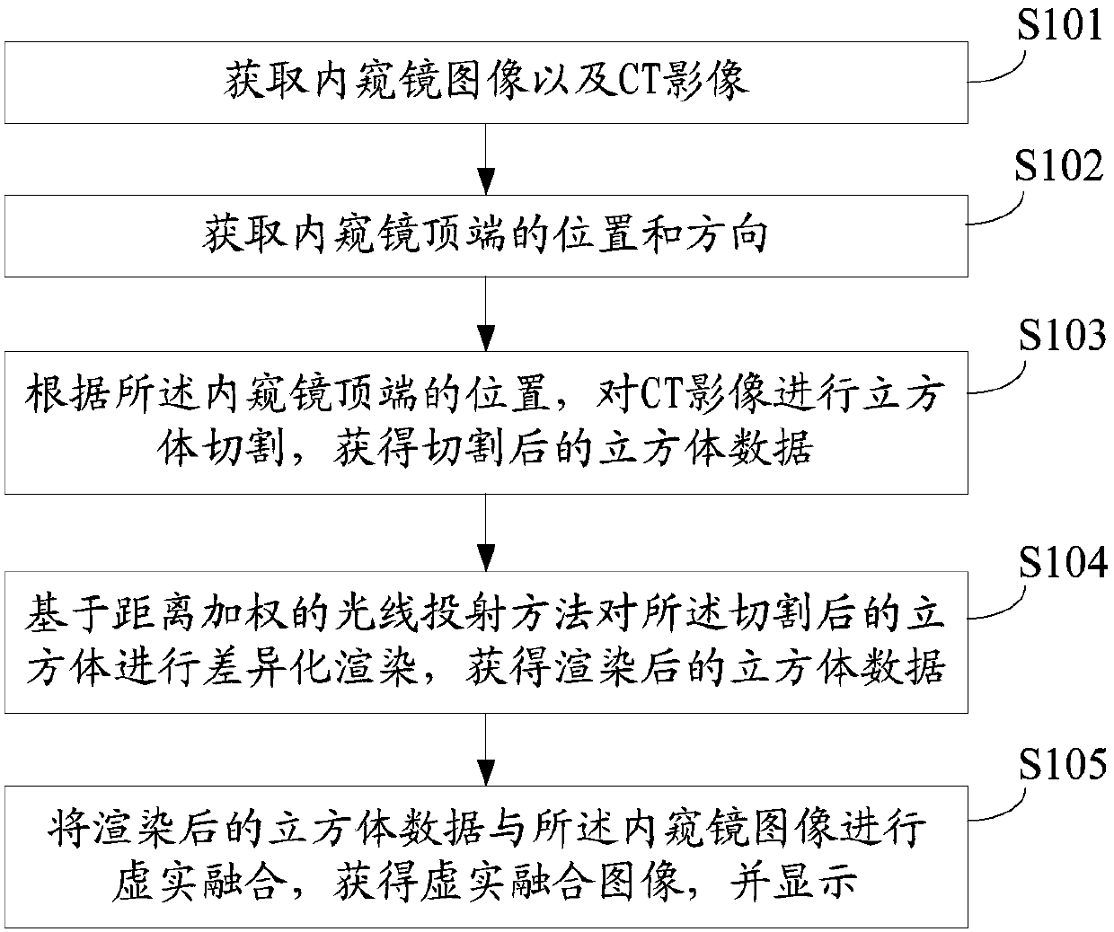
[0049] The embodiment of the present invention provides an image display method in the navigation process of endoscopic minimally invasive surgery. The endoscopic minimally invasive surgery includes, but is not limited to, nose and sinus malignant tumor surgery, skull base tumor surgery, etc. Includes other endoscopic procedures.

[0050] Spe...


PUM


Abstract

The invention discloses an image display method, a device and a system for transanal endoscopic microsurgery navigation. The image display method comprises the following steps of acquiring a CT imageand acquiring an endoscope image in real time; acquiring the position and the direction of the top end of an endoscope in real time; according to the position and the direction of the top end of the endoscope, performing the cube cutting on the CT image to obtain cut cube data; based on a distance-weighted light projection method, subjecting the cut cube data to differential rendering to obtain rendered cube data; subjecting the rendered cube data and the endoscope image to virtual-real fusion treatment to obtain a virtual-real fusion image; and displaying the image. The image display device and the system adopt the above image display method, wherein the image display in the operation navigation process is achieved.

Description

technical field [0001] The invention relates to the field of medical technology, in particular to an image display method, device and system for endoscopic minimally invasive surgery navigation. Background technique [0002] Due to the deep location of skull base tumors and the complex and difficult to distinguish adjacent structures, and the diagnosis and treatment process involves multidisciplinary techniques such as neurosurgery, otolaryngology, and head and neck surgery, it is difficult to completely remove the tumor. After more than a hundred years of development, the diagnosis and treatment of skull base tumors has evolved from naked-eye craniotomy to endoscopic minimally invasive surgery. The endoscopic minimally invasive technique is simple in operation and quick in postoperative recovery. Guided by endoscopic images, it can avoid the damage of the surgical approach to the facial skin structure, thereby reducing the probability of various complications. [0003] At ...

Claims


Application Information

Patent Timeline
IPC IPC(8): G06T7/00G06T7/11G06T7/30G06T7/70G06T5/00G06T5/50
Inventor 杨峰
Owner ARIEMEDI MEDICAL SCI BEIJING CO LTD
Why Eureka
Try Eureka