
Denoising system of medical x-ray image

An optical image and medical technology, applied in the medical field, can solve the problems of sensitivity, poor image oscillation method, no mixed noise, etc., to improve image quality, protect image edge detail information, and achieve the effect of denoising

Active Publication Date: 2018-02-16
SHENZHEN BASDA MEDICAL APP
View PDF2 Cites 14 Cited by
  • Summary
  • Abstract
  • Description
  • Claims
  • Application Information

AI Technical Summary

Problems solved by technology

Because X-ray images generally produce mixed noise, including non-Gaussian and Gaussian parts, none of the above methods can effectively filter out the mixed noise part
[0004] Invention Patent Application No. 200710070013.6 proposes a medical image segmentation method based on an oscillatory network, which is aimed at problems such as difficulty in segmenting images with uneven illumination with a unified threshold, and sensitivity to noise and uneven gray levels.
Combines the advantages of the edge tracking-based method and the region-growing method in image segmentation, but the image oscillation method with blurred boundaries is not effective
[0005] The invention patent CN103500441A only regards the noise as Gaussian noise when processing X-ray images, ignoring the non-Gaussian part, which makes the method have certain limitations when used

Method used


Image

  • Denoising system of medical x-ray image
  • Denoising system of medical x-ray image
  • Denoising system of medical x-ray image

Examples


Embodiment 1

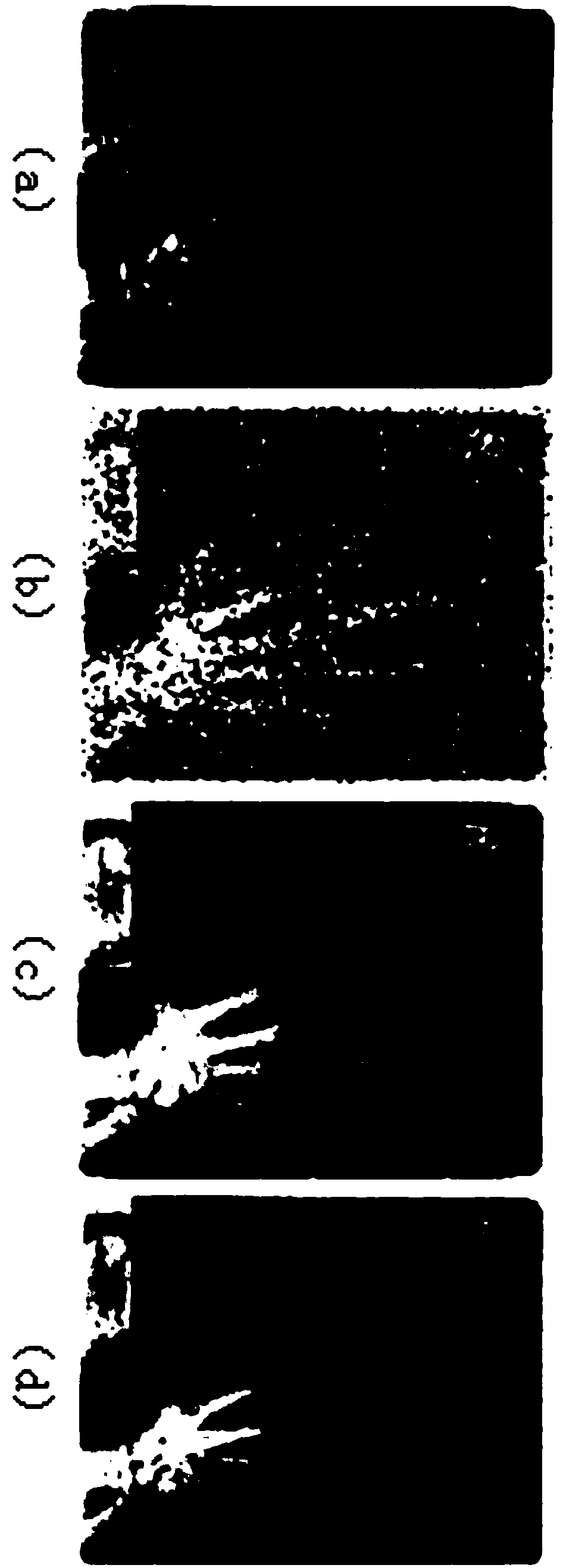
[0026] During the collection, processing and transmission of medical X-ray images, due to the influence of various reasons, there will be more or less various noises and poor contrast. Among them, the type of noise is also relatively complicated. For the convenience of research, it can be divided into two parts: Gaussian noise and non-Gaussian noise. Medically, the requirements for image clarity are relatively high, so looking for a method that can eliminate two types of noise at the same time Yes X The key to optical image denoising. The non-Gaussian part here is mainly salt and pepper noise, and the Gaussian part is mainly granular Gaussian noise. When the decomposition module is processed, the non-Gaussian noise is mainly salt and pepper noise.

[0027] Since the non-Gaussian noise is randomly distributed h 1 (x)=(x 1 ,x 2 ,x 3 ,x 4 ,...,x n ), x i (i=1,2.3...n) are decomposed image pixels, and the arrangement of each noise point is irregular. When denoising, it wil...

Embodiment 2

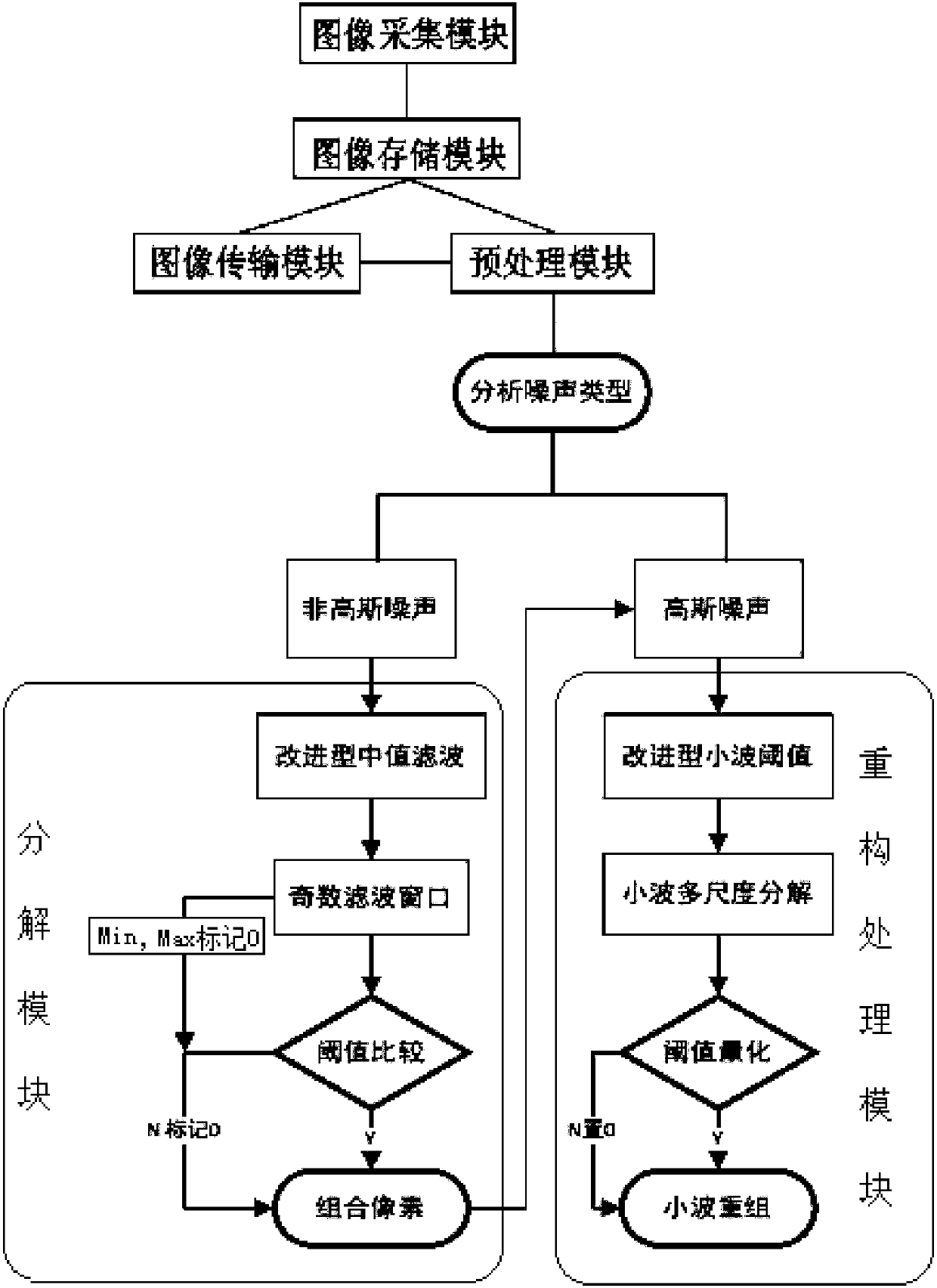
[0031] When the decomposition module is processing, the decomposition module uses the improved median filter to filter out the non-Gaussian part of the noisy image. The specific operation method is:

[0032] a. Using a 3×3 odd-numbered template, within the filtering window, the target pixel H(i, j), and G(i, j) after median filtering;

[0033] b. MAX(H(i, j)) & MIN(H(i, j)) in the window are noise points, marked as 0, others are signal points, marked as 1. filter window such as figure 2 shown;

[0034] c. Observe λ i the value of (i=1, 2, 3, ..., 6);

[0035] where: λ 1 =2×H(i,j)-H(i-1,j)-H(i+1,j),λ 2 =2×H(i, j)-H(i, j-1)-H(i, j+1), λ 3 =2H(i,j)-H(i-1,j-1)-H(i+1,j+1),λ 4 =2H(i,j)-H(i+1,j-1)-H(i-1,j+1);

[0036] H(i-1, j-1)

H(i-1,j)

H(i-1, j+1)

H(i, j-1)

H(i,j)

H(i,j+1)

H(i+1, j-1)

H(i+1,j)

H(i+1, j+1)

[0037] select lambda 1 ~λ 4 average of Compared with the threshold T, when it is greater than or equal to the th...

Embodiment 3

[0042] When the reconstruction processing module is processed, the Gaussian noise is mainly granular Gaussian noise; because the traditional thresholding method is mainly hard thresholding and soft thresholding. The discontinuity of the hard threshold will cause a large mean square error and oscillation in the processed image. There is always a constant deviation between the wavelet coefficients estimated by soft threshold and the actual wavelet coefficients, which makes the processed image too smooth, especially the boundary of the image. The invention uses a new improved threshold value method, which makes up for the shortage of hard threshold value and soft threshold value, and better protects the edge information of the image.

[0043] The reconstruction processing module uses the improved wavelet threshold method to filter out Gaussian noise. The specific operation method is: use the time-frequency characteristics and multi-resolution of wavelet to decompose the noisy ima...


PUM


Abstract

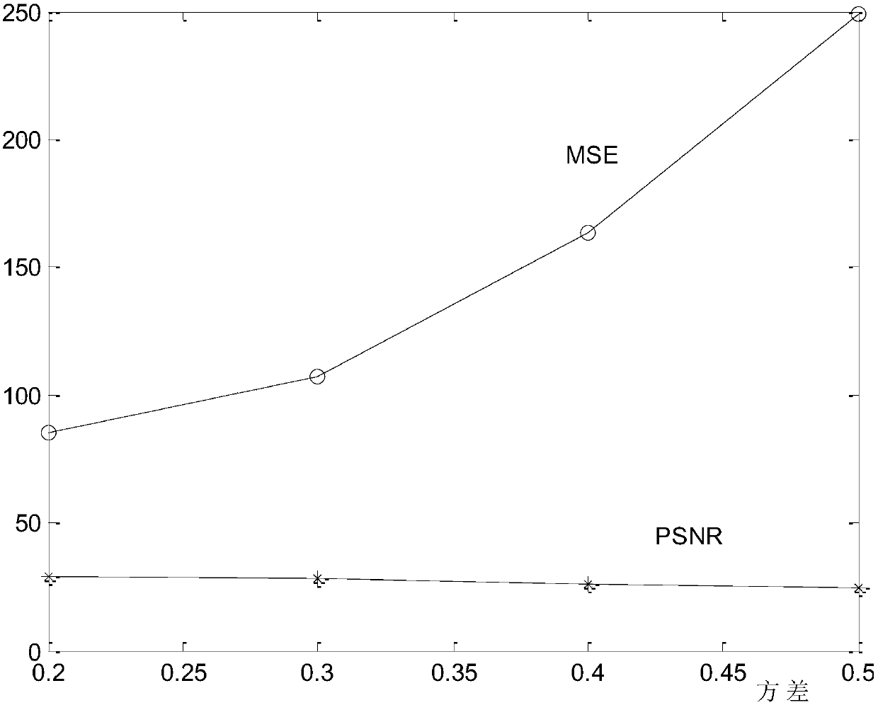
The invention belongs to the field of medical science, and particularly relates to a denoising system of a medical x-ray image. The system comprises a preprocessing module, a decomposition module anda reconstruction processing module which are connected end to end in sequence. The preprocessing module is further connected with an acquisition module, an image storage module and an image transmission module. The image acquisition module is connected with the image storage module. The image storage module is connected with the preprocessing module and the image transmission module. The image transmission module is connected with the preprocessing module. The denoising system of the medical x-ray image can be compiled into operation codes and stored into an x computer hard disk. During the x-ray image acquisition process, the system is automatically operated, so that the denoising effect of x-ray images is effectively achieved. The defects that a hard threshold is not continuous and a soft threshold value has constant deviation can be overcome by adopting the improved wavelet threshold method. The local features of the wavelet can effectively protect the edge detail information of images. Therefore, the system is relatively suitable for noises with relatively small noise variance.

Description

technical field [0001] The invention belongs to the field of medicine, in particular to a denoising system for medical X-ray images. Background technique [0002] X-ray medical images are an important reference for modern medical diagnosis of diseases. Rays are the information source of X-ray images. They were first discovered by the famous German physicist Roentgen in 1895 and later used in medical imaging. The imaging principle of X-ray images is that due to the different densities of the components of the object to be imaged, the absorption of X-rays is also different, and the intensity of transmitted X-rays is different, thus forming an X-ray image on the latex. This technology allows the patient's internal conditions to be observed by the doctor without surgery, reducing the pain of the patient. With the development of computer technology, X-ray digital images have been realized, which is more convenient for storage and transmission. During the imaging process of X-ra...

Claims


Application Information

Patent Timeline
Patent Type & Authority Applications(China)
IPC IPC(8): G06T5/00G06T5/10G06T7/136
CPCG06T5/10G06T7/136G06T2207/10081G06T2207/30008G06T5/70
Inventor 陈值英侯小冉汪红燕
Owner SHENZHEN BASDA MEDICAL APP
Learn More